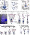
Linking the Primary Cilium to Cell Migration in Tissue Repair and Brain Development
- PMID: 26955067
- PMCID: PMC4776690
- DOI: 10.1093/biosci/biu179
Linking the Primary Cilium to Cell Migration in Tissue Repair and Brain Development
Abstract
Primary cilia are unique sensory organelles that coordinate cellular signaling networks in vertebrates. Inevitably, defects in the formation or function of primary cilia lead to imbalanced regulation of cellular processes that causes multisystemic disorders and diseases, commonly known as ciliopathies. Mounting evidence has demonstrated that primary cilia coordinate multiple activities that are required for cell migration, which, when they are aberrantly regulated, lead to defects in organogenesis and tissue repair, as well as metastasis of tumors. Here, we present an overview on how primary cilia may contribute to the regulation of the cellular signaling pathways that control cyclic processes in directional cell migration.
Keywords: cell migration; cell polarity; cellular signaling; development; primary cilia; tissue repair.
Figures


Similar articles
-
The primary cilium: guardian of organ development and homeostasis.Organogenesis. 2014 Jan 1;10(1):62-8. doi: 10.4161/org.28910. Epub 2014 Apr 17. Organogenesis. 2014. PMID: 24743231 Free PMC article. Review.
-
Cilia and coordination of signaling networks during heart development.Organogenesis. 2014 Jan 1;10(1):108-25. doi: 10.4161/org.27483. Epub 2013 Dec 17. Organogenesis. 2014. PMID: 24345806 Free PMC article. Review.
-
The primary cilium coordinates signaling pathways in cell cycle control and migration during development and tissue repair.Curr Top Dev Biol. 2008;85:261-301. doi: 10.1016/S0070-2153(08)00810-7. Curr Top Dev Biol. 2008. PMID: 19147009 Review.
-
Signaling through the Primary Cilium.Front Cell Dev Biol. 2018 Feb 8;6:8. doi: 10.3389/fcell.2018.00008. eCollection 2018. Front Cell Dev Biol. 2018. PMID: 29473038 Free PMC article. Review.
-
Regulation of cilium length and intraflagellar transport.Int Rev Cell Mol Biol. 2013;303:101-38. doi: 10.1016/B978-0-12-407697-6.00003-9. Int Rev Cell Mol Biol. 2013. PMID: 23445809 Review.
Cited by
-
GJA1 depletion causes ciliary defects by affecting Rab11 trafficking to the ciliary base.Elife. 2022 Aug 25;11:e81016. doi: 10.7554/eLife.81016. Elife. 2022. PMID: 36004726 Free PMC article.
-
Endothelial dysfunction in pulmonary arterial hypertension: loss of cilia length regulation upon cytokine stimulation.Pulm Circ. 2018 Apr-Jun;8(2):2045894018764629. doi: 10.1177/2045894018764629. Epub 2018 Feb 26. Pulm Circ. 2018. PMID: 29480152 Free PMC article.
-
Compartmentalized ciliation changes of oligodendrocytes in aged mouse optic nerve.J Neurosci Res. 2024 Jan;102(1):e25273. doi: 10.1002/jnr.25273. J Neurosci Res. 2024. PMID: 38284846 Free PMC article.
-
Deficiency of the Planar Cell Polarity Protein Intu Delays Kidney Repair and Suppresses Renal Fibrosis after Acute Kidney Injury.Am J Pathol. 2023 Mar;193(3):275-285. doi: 10.1016/j.ajpath.2022.12.006. Epub 2022 Dec 28. Am J Pathol. 2023. PMID: 36586478 Free PMC article.
-
Focal adhesion-related non-ciliary functions of CEP290.PLoS One. 2025 Jul 9;20(7):e0325921. doi: 10.1371/journal.pone.0325921. eCollection 2025. PLoS One. 2025. PMID: 40632733 Free PMC article.
References
-
- Albrecht-Buehler G. Phagokinetic tracks of 3T3 cells: Parallels between the orientation of track segments and of cellular structures which contain actin or tubulin. Cell. 1977;12:333–339. - PubMed
-
- Amin N, Vincan E. The Wnt signaling pathways and cell adhesion. Frontiers in Bioscience. 2012;17:784–804. - PubMed
-
- Ayala R, Shu T, Tsai LH. Trekking across the brain: The journey of neuronal migration. Cell. 2007;128:29–43. - PubMed
-
- Barker AR, McIntosh K, Dawe HR. Meckel–Gruber syndrome proteins are required for actin cytoskeleton organisation and directional cell migration. Molecular Biology of the Cell. 2013;24:24. A442.
LinkOut - more resources
Full Text Sources
Other Literature Sources

