. 2015 Aug 1;65(8):783-797.
doi: 10.1093/biosci/biv084. Epub 2015 Jun 26.

Extracellular Vesicles: Composition, Biological Relevance, and Methods of Study

Affiliations

Extracellular Vesicles: Composition, Biological Relevance, and Methods of Study

MikoŁaj P Zaborowski et al. Bioscience. .

Abstract

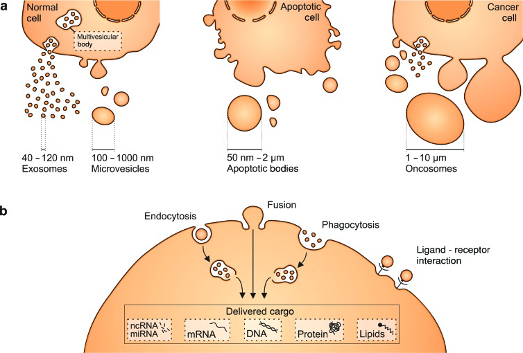
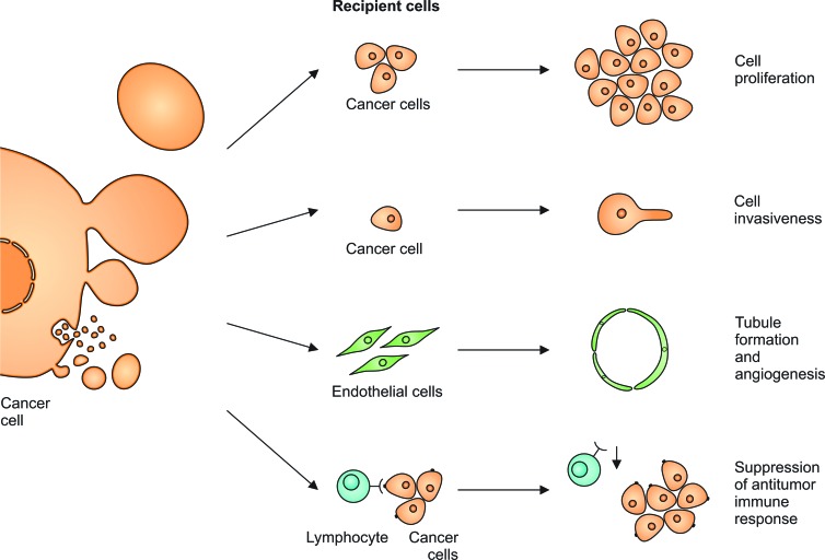
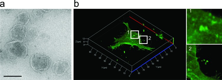
The release of extracellular vesicles (EVs), including exosomes and microvesicles, is a phenomenon shared by many cell types as a means of communicating with other cells and also potentially removing cell contents. The cargo of EVs includes the proteins, lipids, nucleic acids, and membrane receptors of the cells from which they originate. EVs released into the extracellular space can enter body fluids and potentially reach distant tissues. Once taken up by neighboring and/or distal cells, EVs can transfer functional cargo that may alter the status of recipient cells, thereby contributing to both physiological and pathological processes. In this article, we will focus on EV composition, mechanisms of uptake, and their biological effects on recipient cells. We will also discuss established and recently developed methods used to study EVs, including isolation, quantification, labeling and imaging protocols, as well as RNA analysis.

Keywords: exosomes; extracellular vesicles; intercellular communication; methods; microvesicles.


Figures

Figure 1.
Figure 1.
Cells produce different types of extracellular vesicles (EVs) that vary in size. (a) Exosomes and microvesicles (MVs) are produced by normal and diseased cells. Apoptosis triggers the release of apoptotic bodies. In addition, some cancer cells were reported to generate large EVs, termed oncosomes. (b) EVs can be taken up via different mechanisms, including endocytosis, membrane fusion, or phagocytosis. They deliver nucleic acids, proteins, and lipids that can be functional in recipient cells. Ligand–receptor interactions on the cell surface can also result in biological effects and help to target vesicles to specific cell types. Abbreviations: nm, nanometers; μm, micrometers.
Figure 2.
Figure 2.
Extracellular vesicles (EVs) released from transformed cells can exert biological effects. They increase the proliferation rate and invasiveness of other cancer cells. They stimulate endothelial cells to form tubules that support tumor angiogenesis. Tumor-derived EVs render cytotoxic T lymphocytes less reactive, which results in the suppression of antitumor immune response.
Figure 3.
Figure 3.
A visualization of extracellular vesicles. (a) A transmission electron micrograph showing extracellular vesicles (EVs) isolated from HEK293T cells. Note the lipid bilayer–enclosed vesicle characteristic of EVs. Bar, 100 nanometers. 
(b) Live-cell confocal microscopy of human embryonic kidney (HEK) 293T cells expressing palmitoylated GFP (PalmGFP). Plasma membranes are labeled with PalmGFP, allowing the observation of bud-like structures on the cell surface (subpanel 1), suggesting their subsequent release as EVs. Released PalmGFP-EVs were readily observed around the microenvironment of 293T-PalmGFP cells (subpanel 2).

References

    1. Aga M, Bentz GL, Raffa S, Torrisi MR, Kondo S, Wakisaka N, Yoshizaki T, Pagano JS, Shackelford J. Exosomal HIF1α supports invasive potential of nasopharyngeal carcinoma-associated LMP1-positive exosomes. Oncogene. 2014;33:4613–4622. - PMC - PubMed
    1. Al-Nedawi K, Meehan B, Kerbel RS, Allison AC, Rak J. Endothelial expression of autocrine VEGF upon the uptake of tumor-derived microvesicles containing oncogenic EGFR; Proceedings of the National Academy of Sciences; 2009. pp. 3794–3799. - PMC - PubMed
    1. Al-Nedawi K, Meehan B, Micallef J, Lhotak V, May L, Guha A, Rak J. Intercellular transfer of the oncogenic receptor EGFRvIII by microvesicles derived from tumour cells. Nature Cell Biology. 2008;10:619–624. - PubMed
    1. Antonyak MA, Li B, Boroughs LK, Johnson JL, Druso JE, Bryant KL, Holowka DA, Cerione RA. Cancer cell–derived microvesicles induce transformation by transferring tissue transglutaminase and fibronectin to recipient cells; Proceedings of the National Academy of Sciences; 2011. pp. 4852–4857. - PMC - PubMed
    1. Arenaccio C, Chiozzini C, Columba-Cabezas S, Manfredi F, Affabris E, Baur A, Federico M. Exosomes from human immunodeficiency virus type 1 (HIV-1)–infected cells license quiescent CD4+ T lymphocytes to replicate HIV-1 through a Nef- and ADAM17-dependent mechanism. Journal of Virology. 2014;88:11529–11539. - PMC - PubMed