. 2015 Sep:82:23-74.
doi: 10.1016/j.simyco.2015.10.001. Epub 2015 Nov 18.

Common but different: The expanding realm of Cladosporium

Affiliations

Common but different: The expanding realm of Cladosporium

K Bensch et al. Stud Mycol. 2015 Sep.

Abstract

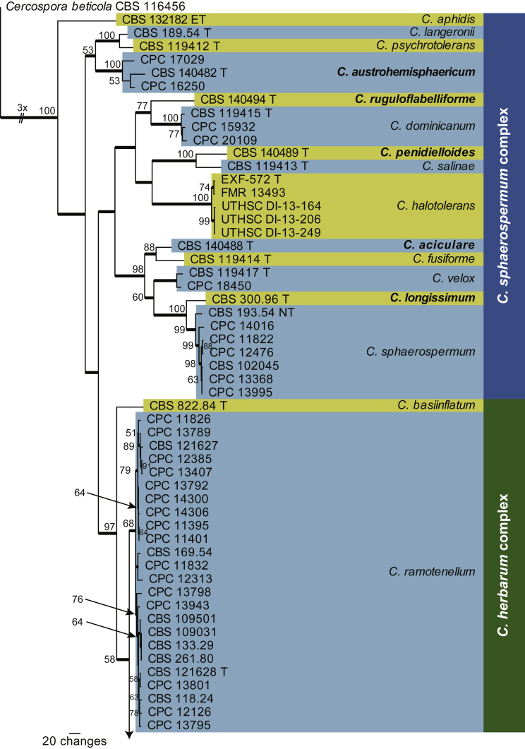
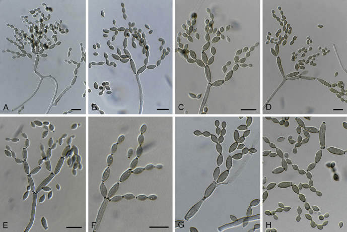
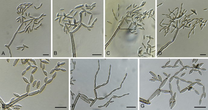
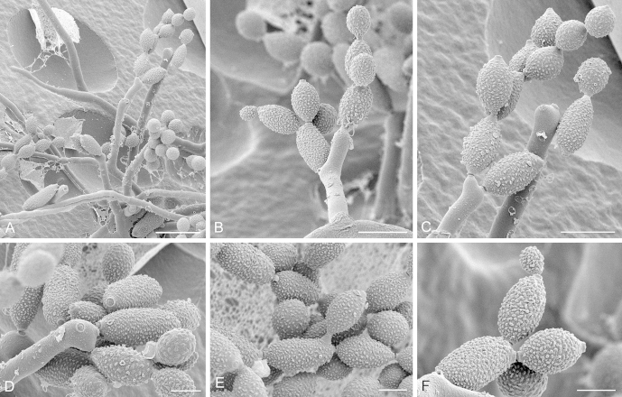
The genus Cladosporium (Cladosporiaceae, Dothideomycetes), which represents one of the largest genera of dematiaceous hyphomycetes, has been intensively investigated during the past decade. In the process, three major species complexes (C. cladosporioides, C. herbarum and C. sphaerospermum) were resolved based on morphology and DNA phylogeny, and a monographic revision of the genus (s. lat.) published reflecting the current taxonomic status quo. In the present study a further 19 new species are described based on phylogenetic characters (nuclear ribosomal RNA gene operon, including the internal transcribed spacer regions ITS1 and ITS2, as well as partial actin and translation elongation factor 1-α gene sequences) and morphological differences. For a selection of the species with ornamented conidia, scanning electron microscopic photos were prepared to illustrate the different types of surface ornamentation. Surprisingly, during this study Cladosporium ramotenellum was found to be a quite common saprobic species, being widely distributed and occurring on various substrates. Therefore, an emended species description is provided. Furthermore, the host range and distribution data for several previously described species are also expanded.

Keywords: C. aggregatocicatricatum Bensch, Crous & U. Braun; C. angustiherbarum Bensch, Crous & U. Braun; C. angustiterminale Bensch, Crous & U. Braun; C. austroafricanum Bensch, Crous & U. Braun; C. austrohemisphaericum Bensch, Crous & U. Braun; C. ipereniae Bensch, Crous & U. Braun; C. limoniforme Bensch, Crous & U. Braun; C. longicatenatum Bensch, Crous & U. Braun; C. longissimum Bensch, Crous & U. Braun; C. montecillanum Bensch, Crous & U. Braun; C. parapenidielloides Bensch, Crous & U. Braun; C. penidielloides Bensch, Crous & U. Braun; C. pseudochalastosporoides Bensch, Crous & U. Braun; C. puyae Bensch, Crous & U. Braun; C. rhusicola Bensch, Crous & U. Braun; C. ruguloflabelliforme Bensch, Crous & U. Braun; C. rugulovarians Bensch, Crous & U. Braun; C. versiforme Bensch, Crous & U. Braun; Cladosporiaceae; Cladosporium aciculare Bensch, Crous & U. Braun; Emendation; Phylogeny; Taxonomic novelties; Taxonomy.


Figures

Fig. 1
Fig. 1
The first of 96 equally most parsimonious trees obtained from a heuristic search with 100 random taxon additions of the combined ITS, tef1 and act sequence alignment of the cladosporioides complex using PAUP v. 4.0b10. The scale bar shows 20 changes, and bootstrap support values from 1 000 replicates are shown at the nodes. Thickened lines indicate the strict consensus branches and species names are indicated to the right of the tree. Species boundaries are indicated with coloured blocks. Names of novel species are printed in bold face and the type status of strains are indicated next to the culture collection number (T: ex-type; ET: ex-epitype; NT: ex-neotype; R: reference). The tree was rooted to Cercospora beticola (CBS 116456).
Fig. 1
Fig. 1
The first of 96 equally most parsimonious trees obtained from a heuristic search with 100 random taxon additions of the combined ITS, tef1 and act sequence alignment of the cladosporioides complex using PAUP v. 4.0b10. The scale bar shows 20 changes, and bootstrap support values from 1 000 replicates are shown at the nodes. Thickened lines indicate the strict consensus branches and species names are indicated to the right of the tree. Species boundaries are indicated with coloured blocks. Names of novel species are printed in bold face and the type status of strains are indicated next to the culture collection number (T: ex-type; ET: ex-epitype; NT: ex-neotype; R: reference). The tree was rooted to Cercospora beticola (CBS 116456).
Fig. 2
Fig. 2
The first of 1 000 equally most parsimonious trees obtained from a heuristic search with 100 random taxon additions of the combined ITS, tef1 and act sequence alignment of the herbarum / sphaerospermum complexes using PAUP v. 4.0b10. The scale bar shows 20 changes, and bootstrap support values from 1 000 replicates are shown at the nodes. Thickened lines indicate the strict consensus branches and species names are indicated to the right of the tree. Species boundaries are indicated with coloured blocks. Names of novel species are printed in bold face and the type status of strains are indicated next to the culture collection number (T: ex-type; ET: ex-epitype; NT: ex-neotype; R: reference). The tree was rooted to Cercospora beticola (CBS 116456).
Fig. 2
Fig. 2
The first of 1 000 equally most parsimonious trees obtained from a heuristic search with 100 random taxon additions of the combined ITS, tef1 and act sequence alignment of the herbarum / sphaerospermum complexes using PAUP v. 4.0b10. The scale bar shows 20 changes, and bootstrap support values from 1 000 replicates are shown at the nodes. Thickened lines indicate the strict consensus branches and species names are indicated to the right of the tree. Species boundaries are indicated with coloured blocks. Names of novel species are printed in bold face and the type status of strains are indicated next to the culture collection number (T: ex-type; ET: ex-epitype; NT: ex-neotype; R: reference). The tree was rooted to Cercospora beticola (CBS 116456).
Fig. 3
Fig. 3
Cladosporium aciculare (CBS 140488). A–C. Conidiophores and conidial chains. D–F. Tip of conidiophores and numerous conidia. G. Ramoconidium and conidia. Scale bars = 10 μm.
Fig. 4
Fig. 4
Cladosporium aggregatocicatricatum (CBS 140493). A–B, D, F. Conidiophores and conidia. C, E. Macronematous conidiophores with conidiogenous loci situated at about the same level on lateral prolongations or round about the stalk at about the same height. G. Tip of a conidiophore with several conidiogenous loci forming conidia. H. Conidia. Scale bars = 10 μm.
Fig. 5
Fig. 5
Cladosporium aggregatocicatricatum (CBS 140493). A. Part of a colony showing substrate hyphae, aerial hyphae and sparse elongated conidiophores. B–D. Conidiophores with conidial chains. Note the highly elongated secondary ramoconidia in (B) with lack of ornamentation at the connection of conidia. Note also the shape of the conidiophore in (C). E. Hila on a secondary ramoconidium and conidiogenous loci on geniculate conidiophores. These areas appear very smooth. F. Two different stages of conidia formation on a secondary ramoconidium. The height of the individual ornamentations is markedly visible here. In (B) and (F) wart-like structures are visible on relatively broad aerial hyphae. Scale bars = 5 (E–F), 10 (B–D), 20 (A) μm.
Fig. 6
Fig. 6
Cladosporium angustiherbarum (CBS 140479). A–G, I. Conidiophores and conidial chains. H. Tip of a conidiophore and ornamented conidia. Scale bars = 10 μm.
Fig. 7
Fig. 7
Cladosporium angustiherbarum (CBS 140479). A. View on the agar surface showing conidiophores sprouting from structures beneath the agar surface or arising from aerial hyphae with conidial chains. B. Running hyphae with a number of conidia. Note the visibility of septa in these hyphae. Behind the septa, initials of conidiophores can be observed as bulges. C. A bundle of aerial hyphae bearing a number of conidiophores that stick out in different directions. D. Netting of aerial hyphae with conidiophores. E, H. Conidiophores with conidial chains. F–G. Conidial chains showing details of surface ornamentation, especially in (G). Ornamentation is present early during the formation of the conidia (F) I. Tip of a capitate conidiophore with conidial chains. I. Secondary ramoconidia with details of conidial hila (including one conidium initial). Scale bars = 2 (G), 5 (E–F, I–J), 10 (B–D, H), 20 (A) μm.
Fig. 8
Fig. 8
Cladosporium angustiterminale (CBS 140480). A–F. Conidiophores and conidial chains. G. Peculiar conidiogenesis characterised by forming several conidiogenous loci at about the same level, followed by continuing growth with narrower conidiophores and additional loci at a higher level. Scale bars = 10 μm.
Fig. 9
Fig. 9
Cladosporium austroafricanum (CBS 140481). A–F. Conidiophores and conidial chains. G. Conidia. Scale bars = 10 μm.
Fig. 10
Fig. 10
Cladosporium austrohemisphaericum (CBS 140482). A–F. Micro-, semimacro- and macronematous conidiophores and conidial chains. G. Ramoconidium and conidia. H. Conidia. Scale bars = 10 μm.
Fig. 11
Fig. 11
Cladosporium ipereniae (CBS 140483). A–D. Unbranched or branched macronematous conidiophores and conidial chains. E–G. Tip of conidiophores and conidia with variable surface ornamentation. Scale bars = 10 μm.
Fig. 12
Fig. 12
Cladosporium ipereniae (CBS 140483). A. Overview of conidiophores and hyphae. B, D. Conidiophores arising from plagiotropous hyphae or sprouting from structures beneath the agar surface with conidial chains. C. Branched conidiophore arising from a plagiotropous hypha with conidial chains. E–F. Tip of conidiophores with conidial chains. Note the constricted septum of the conidiophore in (E). G. Branchlet of a conidiophore with conidia. Note the round shape of the conidia and the elevated scar region. Note also the very short conidiophorous structure on the aerial hyphae. H–I. Conidial chains showing details of the characteristic surface ornamentation that appears to run parallel on the cell wall of larger spores. Note also the scar on the ramoconidium. Scale bars = 2 (H–I), 5 (F–G), 10 (B–C, E), 20 (D), 100 (A) μm.
Fig. 13
Fig. 13
Cladosporium limoniforme (CBS 140484). A–E. Micronematous conidiophores forming large amounts of conidia. F–H. Conidial chains. Scale bars = 10 μm.
Fig. 14
Fig. 14
Cladosporium limoniforme (CBS 140484). A. Overview of a cluster of conidiophores that seem to originate from one base, illustrating the density of conidial chains. B–C, E. Conidiophores and conidial chains either sprouting from structures beneath the agar surface or arising from running hyphae with conidial chains. Note the smooth surface of the conidiophores in contrast to the ornamented conidia (C). Note also the branching conidiophore in (E). D, F. Tip of conidiophores and conidia with details of scars. G–H. Details of conidia, ornamentation and scars. Scale bars = 1 (F), 2 (D, G, H), 10 (B, C, E), 20 (A) μm.
Fig. 15
Fig. 15
Cladosporium longicatenatum (CBS 140485). A, C–E. Conidiophores with long, dichotomously branched conidial chains. B. Ramoconidium with conidial chains. F–G. Tip of conidiophores with conidia attached. Scale bars = 10 μm.
Fig. 16
Fig. 16
Cladosporium longicatenatum (CBS 140485). A. Overview of hyphae and clusters of rounded cells visible on the agar surface that give rise to slender elongated conidiophores and conidia. B. Very elongated smooth secondary ramoconidia give rise to intercalary and small terminal conidia. Note the pattern of secondary ramoconidia on the conidiophore. C. Detail of conidial hila. D–F. Conidiophores, secondary ramoconidia and intercalary conidia from different angles. G–H. Secondary ramoconidia, intercalary and small terminal conidia. Note an aerial hypha in (G) with septa. Scale bars = 2 (C), 5 (E–H), 10 (B, D), 20 (A) μm.
Fig. 17
Fig. 17
Cladosporium longissimum (CBS 300.96). A–C. Macronematous conidiophores and conidial chains. D. Branchlet of a conidiophore and conidia. E. Tip of a conidiophore and conidia. Scale bars = 10 μm.
Fig. 18
Fig. 18
Cladosporium montecillanum (CBS 140486). A–G. Conidiophores and conidial chains. Scale bars = 10 μm.
Fig. 19
Fig. 19
Cladosporium myrtacearum (CPC 16319). A–C. Conidiophores and conidial chains. D–E. Tips of conidiophores and conidia. Scale bars = 10 μm.
Fig. 20
Fig. 20
Cladosporium myrtacearum (CPC 16319). A. Overview on agar surface with conidiophores arising from the surface. B. Running hyphae giving rise to several conidiophores. C–F. Tips of conidiophores with conidial chains. Note that the reticulate surface ornamentation differs among the secondary ramo-, intercalary and terminal conidia. Mostly secondary ramoconidia have less surface ornamentation. G. Details of intercalary and small terminal conidia and hila on conidia. Note the smoother surface of these terminal conidia as if the ornamentation is still being formed. H. Secondary ramoconidium with a remarkable expanded non-ornamented hilus region. I. Very early conidia formed on secondary ramoconidia. J–K. Details of terminal chains, hila and branching of secondary ramoconidia on the conidiophore. Scale bars = 5 (F–K), 10 (C–E), 20 (B), 50 (A) μm.
Fig. 21
Fig. 21
Cladosporium parapenidielloides (CBS 140487). A–D. Conidiophores and conidial chains. E–G. Tip of conidiophores and conidia. Scale bars = 10 μm.
Fig. 22
Fig. 22
Cladosporium parapenidielloides (CBS 140487). A, D. Overview of a part of a colony showing a dense field of conidiophores that arise from cells at the level of the agar surface and forming conidial chains. B. Dislodged conidial chains on the agar surface. C, E. Conidial chains with branching at different secondary ramoconidia and intercalary conidia. F–G. Conidiogenous loci on a conidiophore and conidial hila on secondary ramoconidia. Note the very broad contact area (diameter > 2 μm) between secondary ramoconidium and conidiophore in (G). Ornamentation is absent on these conidia, although some pattern formation on the secondary ramoconidium is visible in this figure. Scale bars = 2 (E–F), 5 (C, G), 10 (A–B, D) μm.
Fig. 23
Fig. 23
Cladosporium penidielloides (CBS 140489). A–F. Macro- and micronematous conidiophores and conidial chains. G. Ramoconidium and conidia. Scale bars = 10 μm.
Fig. 24
Fig. 24
Cladosporium penidielloides (CBS 140489). A. Overview on the agar surface also containing slender conidiophores. B–D, F, H. Short and long conidiophores erumpent from structures beneath the agar or arising from plagiotropous hyphae with conidial chains. E. Conidiogenous cell with a cluster of four pronounced loci and secondary ramoconidia. G, I–J. Conidial chains showing details of conidial hila. Ornamentation is missing in most of the conidia, few show some irregular reticulate structures. Note the large scar / contact region between spores in E and G. Also note the conidium initials in (J). Scale bars = 2 (J), 5 (E–F, I–J), 10 (B–D, H), 20 (A) μm.
Fig. 25
Fig. 25
Cladosporium pseudochalastosporoides (CBS 140490). A–E. Conidiophores and conidial chains. F–G. Tip of conidiophores and conidia. Scale bars = 10 μm.
Fig. 26
Fig. 26
Cladosporium puyae (CBS 274.80A). A–G. Conidiophores and conidial chains. Scale bars = 10 μm.
Fig. 27
Fig. 27
Cladosporium puyae (CBS 274.80A). A. Survey of colony development with numerous conidiophores arising from the agar surface. B–C, E. Conidiophores, hyphae and conidial chains. D. Conidial chain showing the characteristic dispersed surface ornamentation. Note that several of the verrucae are irregularly enlarged. F. Details of conidial surface ornamentation and hila. Note the conidium initial. Scale bars = 2 (F), 10 (C–E), 20 (B), 50 (A) μm.
Fig. 28
Fig. 28
Cladosporium rhusicola (CBS 140492). A–D, F–G. Macro-, semimacro- and micronematous conidiophores and conidial chains. E. Conidial chains. Scale bars = 10 μm.
Fig. 29
Fig. 29
Cladosporium rhusicola (CBS 140492). A. Survey of colony development with swollen hyphal cells that give rise to young conidiophores. The ornamentation, most probably of conidia, is also visible. B. Overview on agar surface with several intact conidiophores arising from the surface level and forming conidial chains. C–D. Conidial chains on conidiophores with scars visible in (C). Note the septated aerial hyphae. Note also the relatively thin connection between the conidia and the absence of ornamentation in the hilar region. E. Several conidial chains form a stabilised superstructure. Note the conidium initial. F, I. Young intact conidiophores with three secondary ramoconidia on the tapered smooth conidiophore. G. Details of strong somewhat dispersed ornamentation and the hilar region with scar. H. Branching pattern of conidial chains and conidium initial. J. Details of ornamentation and connection of spores. Scale bars = 2 (J), 5 (F–G, I), 10 (A, C–E, H), 20 (B) μm.
Fig. 30
Fig. 30
Cladosporium ruguloflabelliforme (CBS 140494). A–E. Conidiophores with conidial chains. F. Tip of a conidiophore with dichotomously branched conidial chains. G. Conidial chain. Scale bars = 10 μm.
Fig. 31
Fig. 31
Cladosporium ruguloflabelliforme (CBS 140494). A. Conidiophore showing several secondary ramoconidia with hila. Note the shape and loci on the conidiophore itself. Note also the irregularly reticulate surface ornamentation of conidia. B. Terminal conidia and ornamentation that seems to be less prominent at forming (young) conidia. C. Dislodged conidial chain, conidiophore, aerial and substrate hyphae in the colony. D–G. Conidiophores, terminal, intercalary and secondary ramoconidia, details of scars and notable ornamentation in G. Scale bars = 5 (B, E–G), 10 (A, C–D) μm.
Fig. 32
Fig. 32
Cladosporium rugulovarians (CBS 140495). A–C. Conidiophores with conidial chains. D–E. Branched conidiophores. F–H. Tips of conidiophores, with ramoconidia, secondary ramoconidia and conidia. Scale bars = 10 μm.
Fig. 33
Fig. 33
Cladosporium rugulovarians (CBS 140495). A. Overview of a colony with a fascicle of long conidiophores arising from the agar. B. Conidiophore with dislodged conidia and conidium initial. C. Conidiophores with secondary ramoconidia forming large amounts of globose, subglobose and ovoid intercalary and small terminal conidia with a fine dispersed reticular ornamentation. D. Conidiophore (left) and conidia showing ornamentation and the hilar region. All structures show scars. E. Intercalary and small terminal conidia showing the irregularly reticulate surface ornamentation. Scale bars = 2 (B, D–E), 10 (C), 100 (A) μm.
Fig. 34
Fig. 34
Cladosporium sinuosum (CPC 14000 and CPC 17632). A–G, I–M (CPC 17632). Geniculate-sinuous conidiophores and conidia, either formed solitary or in short chains. H. Conidia. Scale bars = 10 μm.
Fig. 35
Fig. 35
Cladosporium sinuosum (CPC 14000). A. Overview of a part of a colony on SNA, with numerous aerial hyphae. B. Detailed overview with hyphae, aerial hyphae, conidiophores and ornamented conidia. Note the continuous growth of the conidiophore that forms conidial chains as it goes (see also Figure G). C–F. Aerial hyphae, conidiophores and conidia. Note the aerial hyphal bundles with an anastomosis in Fig. E. Note also the shape of the conidiophore in this Figure. The area of contact between the conidia is not characterised by an absence in ornamentation. G–H. Details of conidia and conidiophores. Geniculate conidiophore with a single conidium formed. H. Details of ornamentation. Scale bars = 5 (H), 10 (C–G), 20 (A–B) μm.
Fig. 36
Fig. 36
Cladosporium sinuosum (CPC 17632) A. Overview showing a very dense area of conidium formation. Some aerial hyphae are visible. B–F. Conidial chains on geniculate conidiophores. The shape of conidia and conidiophores is the same as in Fig. 35. Note the conidium initial in (B), and the geniculate growth of the conidiophore in (F). G. Details of conidia, conidiophores and scars. Note the fine ornamentation of the conidiophore. Scale bars = 5 (G), 10 (B–F), 50 (A) μm.
Fig. 37
Fig. 37
Cladosporium versiforme (CBS 140491). A–G. Macro-, semimacro- and micronematous conidiophores and conidial chains. Scale bars = 10 μm.
Fig. 38
Fig. 38
Cladosporium versiforme (CBS 140491). A. Conidiophores sprouting from hyphae or swollen intercalary cells on the agar surface or arising from an aerial hypha. B–D. Conidiophores with a characteristic rounded shape on which relatively narrow loci can be observed. Note the near geniculate shape of a conidiophore in figure (B). Conidi-ogenous loci which are confined to these areas are shown in more detail together with some conidial hila. E. Conidial chains with triangular hilus. F. Detail of the prominent dense ornamentation on conidia. Note the small area on the spore that remains smooth (see also E) and the strong ornamentation of terminal conidia that are still very small. Scale bars = 5 (D–F), 10 (B–C), 20 (A) μm.

References

    1. Allescher A. Einige weniger bekannte Pilze aus den Gewächshäusern des Kgl. Botan. Gartens in München. Hedwigia. 1895;34:215–221.
    1. Aptroot A. Mycosphaerella and its anamorphs: 2. Conspectus of Mycosphaerella. CBS Biodiversity Series. 2006;5:1–231.
    1. Bensch K., Braun U., Groenewald J.Z. The genus Cladosporium. Studies in Mycology. 2012;72:1–401. - PMC - PubMed
    1. Bensch K., Groenewald J.Z., Dijksterhuis J. Species and ecological diversity within the Cladosporium cladosporioides complex (Davidiellaceae, Capnodiales) Studies in Mycology. 2010;67:1–94. - PMC - PubMed
    1. Braun U., Crous P.W., Dugan F.M. Phylogeny and taxonomy of cladosporium-like hyphomycetes, including Davidiella gen. nov., the teleomorph of Cladosporium s.str. Mycological Progress. 2003;2(1):3–18.