Review
. 2016:2016:4621659.
doi: 10.1155/2016/4621659. Epub 2016 Feb 3.

Pathogenesis and Diagnostic Approaches of Avian Infectious Bronchitis

Affiliations
Review

Pathogenesis and Diagnostic Approaches of Avian Infectious Bronchitis

Faruku Bande et al. Adv Virol. 2016.

Abstract

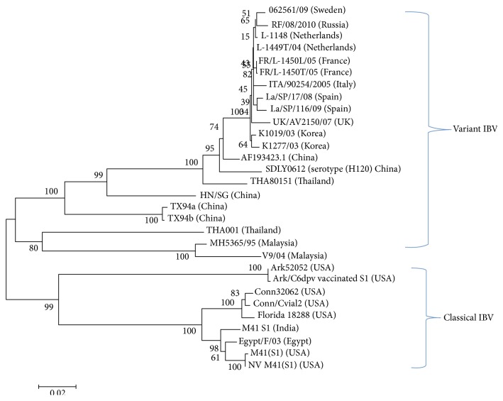
Infectious bronchitis (IB) is one of the major economically important poultry diseases distributed worldwide. It is caused by infectious bronchitis virus (IBV) and affects both galliform and nongalliform birds. Its economic impact includes decreased egg production and poor egg quality in layers, stunted growth, poor carcass weight, and mortality in broiler chickens. Although primarily affecting the respiratory tract, IBV demonstrates a wide range of tissues tropism, including the renal and reproductive systems. Thus, disease outcome may be influenced by the organ or tissue involved as well as pathotypes or strain of the infecting virus. Knowledge on the epidemiology of the prevalent IBV strains in a particular region is therefore important to guide control and preventions. Meanwhile previous diagnostic methods such as serology and virus isolations are less sensitive and time consuming, respectively; current methods, such as reverse transcription polymerase chain reaction (RT-PCR), Restriction Fragment Length Polymorphism (RFLP), and sequencing, offer highly sensitive, rapid, and accurate diagnostic results, thus enabling the genotyping of new viral strains within the shortest possible time. This review discusses aspects on pathogenesis and diagnostic methods for IBV infection.


Figures

Figure 1
Figure 1
Dullness exhibited in chickens infected following experimental infection with IBV (courtesy: Siti Suri Arshad).
Figure 2
Figure 2
Irregularity in the shape and sizes of eggs from natural IBV infected breeder chickens (a). Watery albumen from IBV infected chicken ((b) left) compared to normal egg ((b) right).
Figure 3
Figure 3
Gross lesions observed on respiratory organs of chicken naturally infected with IBV. Presence of mucoid secretion, congestion, and hyperaemia in the trachea (a); mild focal areas of lung consolidation (b).
Figure 4
Figure 4
Histopathological changes in the trachea of naturally IBV infected chicken. Note: the marked infiltration of lymphocytes within the epithelia (black arrow (b)) and evidence of mucosal secretions of goblet cells (yellow arrow (a)).
Figure 5
Figure 5
Gross lesions in kidney of chicken following experimental infection with a nephropathogenic infectious bronchitis virus. Note: swelling and congestion of the kidney (arrow) (courtesy: Siti Suri Arshad).
Figure 6
Figure 6
Chicken showing natural IBV infection. Accumulation of egg yolk in abdominal cavity (a); slightly enlarged, pale, friable liver (b) and multiple petechial haemorrhages on the serosal surfaces of proventriculus (c), gizzard (d), and small intestine (e).
Figure 7
Figure 7
Cystic oviduct in 11-week-old chicken experimentally infected with a CR88 infectious bronchitis virus strain. Note the distention of the entire oviduct and fluid accumulation (arrow).
Figure 8
Figure 8
Embryo development at 17 days old following inoculations with IBV-CR88 strain. Note evidence of dwarfism and curling of the toes in IBV infected embryo (right) compared to a noninfected control embryo (left).
Figure 9
Figure 9
Negative staining electron microscope showing spherical shape of virus with typical spike projections (arrow) surrounding the virion of avian infectious bronchitis virus (courtesy: Siti Suri Arshad).
Figure 10
Figure 10
Electropherogram showing 1.7 kb RT-PCR amplified S1 genes from vaccine (H120) and virulent (M41) IBV strains (a) compared to a 320 bp RT-PCR amplified N-gene (b) of H120 and M41 IBV serotypes. Lane M = 1 kb molecular ladder (a) and 100 bp ladder (b); lane NC: negative control (no template control).
Figure 11
Figure 11
Neighbour joining phylogenetic analysis based on nucleotide acid sequence of S1-spike gene of classical and variant IBV strains identified in different countries. The tree was drawn with MEGA5 software using 1000 bootstrap replicates.

References

    1. Cavanagh D. Coronavirus avian infectious bronchitis virus. Veterinary Research. 2007;38(2):281–297. doi: 10.1051/vetres:2006055. - DOI - PubMed
    1. Arshad S. S. Infectious bronchitis. In: Zamri-Saad M., editor. Diseases of Poultry in South East Asia. Serdang, Malaysia: Universiti Putra Malaysia Press; 2006. pp. 199–206.
    1. Schalk A., Hawn M. An apparently new respiratory disease of baby chicks. Journal of the American Veterinary Medical Association. 1931;78:413–422.
    1. Lai M. M., Cavanagh D. The molecular biology of coronaviruses. Advances in Virus Research. 1997;48:1–100. - PMC - PubMed
    1. de Haan C. A. M., Vennema H., Rottier P. J. M. Assembly of the coronavirus envelope: homotypic interactions between the M proteins. Journal of Virology. 2000;74(11):4967–4978. doi: 10.1128/jvi.74.11.4967-4978.2000. - DOI - PMC - PubMed

LinkOut - more resources