. 2015 Dec 1;10(6):759-776.
doi: 10.1016/j.nantod.2015.11.009.

Nanomedicines for Endothelial Disorders

Affiliations

Nanomedicines for Endothelial Disorders

Bomy Lee Chung et al. Nano Today. .

Abstract

The endothelium lines the internal surfaces of blood and lymphatic vessels and has a critical role in maintaining homeostasis. Endothelial dysfunction is involved in the pathology of many diseases and conditions, including disorders such as diabetes, cardiovascular diseases, and cancer. Given this common etiology in a range of diseases, medicines targeting an impaired endothelium can strengthen the arsenal of therapeutics. Nanomedicine - the application of nanotechnology to healthcare - presents novel opportunities and potential for the treatment of diseases associated with an impaired endothelium. This review discusses therapies currently available for the treatment of these disorders and highlights the application of nanomedicine for the therapy of these major disease complications.

Keywords: atherosclerosis; cancer; diabetes; endothelial disorder; endothelium; nanomedicine; permeability.


Conflict of interest statement

Competing financial interests

R.L. and O.C.F. have financial interests in BIND Therapeutics, Selecta Biosciences, and Blend Therapeutics, which are developing nanoparticle technologies for medical applications. These companies did not support the aforementioned research and currently have no rights to any technology or intellectual property developed as part of this research. All other authors declare no conflicts.

Figures

Figure 1
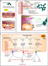
Figure 1. Mechanism and mediators of endothelial function and permeability
(a) Endothelial cells maintain the tight cell-cell connections and the underlying matrix for increased barrier integrity. Sphingosine-1-phosphate (S1P) binds to its EDG-1 receptor, which ultimately strengthens EC barrier function through lamellipodia formation and subsequent AJ assembly. In dysfunctional barriers, thrombin binds to the PAR-1 receptor, which induces inositol trisphosphate (IP3) production and a subsequent increase in intracellular Ca2+. Increased Ca2+ activates the myosin light chain kinase (MLCK) to phosphorylate MLCs, leading to increased actomyosin contractility. Furthermore, thrombin inhibits MLC phosphatase activity through Rho/Rho kinase (RhoK), which increases MLC phosphorylation. The resulting actomyosin contraction contributes to increased permeability of the EC layer. (b) In normal ECs, activated eNOS promotes nitric oxide (NO) production, inhibiting platelet aggregation, leukocyte adhesion, and smooth muscle cell proliferation. Reduced availability of NO leads to endothelial disorder and compromised production of NO by oxidative stress. This inhibits eNOS-derived NO production and results in platelet aggregation and leukocyte adhesion as well as increased contractions of smooth muscle cells.
Figure 2
Figure 2. Timeline of selected clinical stage nanomedicines (FDA-approved and in clinical development)
PEG = polyethylene glycol; siRNA = small interfering RNA; GAH TNF = tumor necrosis factor; Bik = Bcl-2 interacting killer; PEG–PGlu = polyethylene glycol-poly(glutamate); and PEG–PLA = polyethylene glycol-polylactic acid. Phase trial is as of June 2015, in the United States. Source: www.clinicaltrials.gov.
Figure 3
Figure 3. Endothelial disorder in metabolic and cardiovascular diseases
(a) Key EnD inducers and EnD-associated diseases. (b) A key EnD mechanism in diabetes. NO is formed from L-arginine by eNOS. In diabetes characterized by insulin resistance and hyperglycemia, EnD results from reduced production of NO. This arises through decreased activation of eNOS due to insulin resistance and increased breakdown of NO by ROS, promoted by hyperglycemia. (c) Initiation and progression of atherosclerosis with an activated endothelium (adapted from [95]). Atherogenic lipoproteins enter the intima and aggregate within the extracellular intimal space (i). Unregulated uptake of these atherogenic lipoproteins by macrophages leads to the generation of foam cells (ii). In addition to monocytes, other types of leukocyte, particularly T cells, are recruited to atherosclerotic lesions and cause chronic inflammation. The growth of plaque induces tissue remodeling (iii). The foam cells release cellular debris and crystalline cholesterol. Smooth muscle cells form a fibrous cap beneath the endothelium, contributing to the formation of a necrotic core within the plaque. The resulting non-obstructive plaque may rupture, resulting in the formation of a thrombus in the lumen (iv), which can lead to tissue infarction. Ultimately, if the plaque does not rupture and the lesion continues to grow, the lesion can encroach on the lumen and result in clinically obstructive disease (v). Potential NP therapies in atherosclerosis could benefit from the increased microvessel permeability, which is caused by hypoxia-induced neovascularization of the vasa vasorum and would allow the delivery of NPs to plaques within vascular vessel walls.
Figure 4
Figure 4. The role of VEGF in tumor growth and inhibition of VEGF as a cancer therapy
(a) Angiopoietin 1 (ANGPT1) and VEGF play important roles through the actions of circulating VEGF and intracrine actions of endothelial-derived VEGF. (b) Tumors express several pro-angiogenic factors (e.g. VEGF, bFGF, PDGF). Interstitial fibroblasts and dissociated microvascular pericytes, stimulated by PDGF from tumors, release VEGF and contribute to EC proliferation and migration, thereby exerting paracrine EC protective effects during angiogenesis [148]. Blockage of these mechanisms is expected to improve the efficacy of cancer therapy as well as inhibition of pro-angiogenic factors. (c) Strategies to inhibit VEGF signaling include monoclonal antibodies targeting VEGFA, such as Bevacizumab, and VEGF receptors such as IMC-18F1 and Ramucirumab. Also, soluble VEGF receptors, such as VEGF-Trap or VEGLIN, have been used to inhibit VEGF signaling. In ECs, many small-molecule VEGF RTK inhibitors have been tested to prevent ligand-dependent receptor phosphorylation of VEGFR1 and VEGFR2, which would otherwise trigger various signaling pathways, eventually activating angiogenesis [149].
Figure 5
Figure 5. Probing nanoparticle translocation across the permeable endothelium using an in vitro microfluidic model with in vivo validation
(a) Fluorescence microscopy images of typical cross-sections of the healthy wall (L, lumen) and the atherosclerotic vessel with plaque (P). (b) Cross-section schematics of continuous normal and permeable capillaries that penetrate into the atherosclerotic plaque from the vasa vasorum. (c) TEM of the endothelial lining of a normal/healthy vessel wall (left) compared to a permeable endothelial layer with a large gap (right). (Scale bar, 2µm). (d) High resolution TEM showing ECs with a gap between them and a macrophage (MΦ) behind it. At a higher magnification, multiple individual nanoparticles (arrowheads) can be found. (Scale bar, 2µm.) The neovessel (N) within the plaque is bordered with a lipid-loaded MΦ, which itself has taken up nanoparticles as well. (Scale bar, 1µm.) (e) Diagram of an endothelialized microfluidic device enabling TEER measurement across the EC layer. (f) Permeable EC layer with disrupted adherens junctions between ECs, as evidenced by patchy expression of VE-cadherin (green) in the image on the right compared to the left. Nuclei (DAPI). (Scale bar, 20µm). (g) ECs in different culture media show different permeability (shown by TEER and FITC-albumin translocation). (h) Nanoparticle translocation and TEER from these experiments are inversely correlated (r2 = 0.54, P < 0.0001). Reprinted with permission from PNAS [209].

References

    1. Cines DB, Pollak ES, Buck CA, Loscalzo J, Zimmerman GA, McEver RP, Pober JS, Wick TM, Konkle BA, Schwartz BS, Barnathan ES, McCrae KR, Hug BA, Schmidt AM, Stern DM. Blood. 1998;91:3527–3561. - PubMed
    1. Luo BH, Carman CV, Springer TA. Annual review of immunology. 2007;25:619–647. - PMC - PubMed
    1. Mehta D, Malik AB. Physiological reviews. 2006;86:279–367. - PubMed
    1. Bogatcheva NV, Verin AD. Microvascular research. 2008;76:202–207. - PMC - PubMed
    1. Shen Q, Rigor RR, Pivetti CD, Wu MH, Yuan SY. Cardiovascular research. 2010;87:272–280. - PMC - PubMed

LinkOut - more resources