. 2015 Jun 24;1(3):142-147.
doi: 10.1021/acscentsci.5b00121. Epub 2015 Jun 3.

The use of 18F-2-fluorodeoxyglucose (FDG) to label antibody fragments for immuno-PET of pancreatic cancer

Affiliations

The use of 18F-2-fluorodeoxyglucose (FDG) to label antibody fragments for immuno-PET of pancreatic cancer

Mohammad Rashidian et al. ACS Cent Sci. .

Abstract

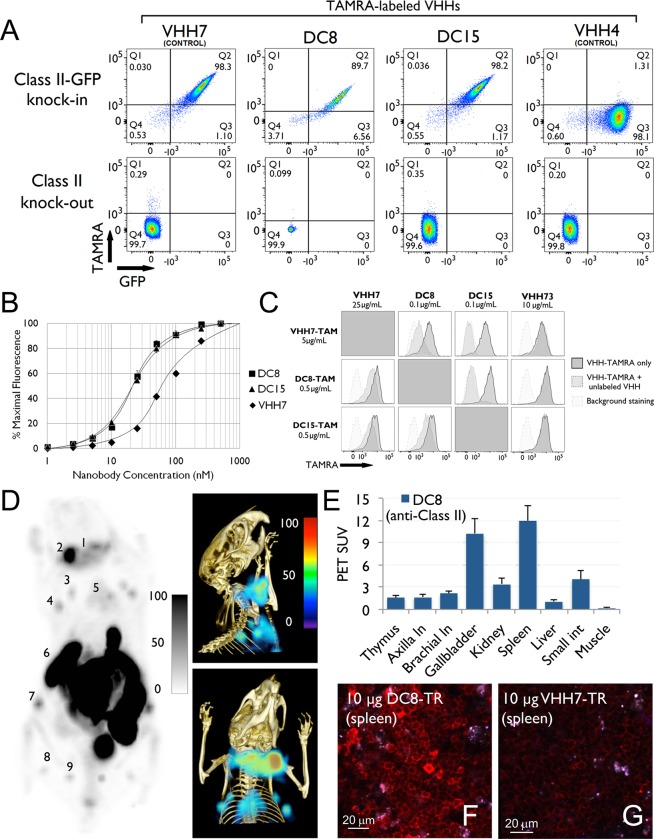
We generated 18F-labeled antibody fragments for PET imaging using a sortase-mediated reaction to install a transcyclooctene (TCO)-functionalized short peptide onto proteins of interest, followed by reaction with a tetrazine-labeled-18F-2-deoxyfluoroglucose (FDG). The method is rapid, robust, and site-specific (radiochemical yields >25%, not decay corrected). The availability of 18F-2-deoxyfluoroglucose avoids the need for more complicated chemistries used to generate carbon-fluorine bonds. We demonstrate the utility of the method by detecting heterotopic pancreatic tumors in mice by PET, using anti-Class II MHC single domain antibodies. We correlate macroscopic PET images with microscopic two-photon visualization of the tumor. Our approach provides easy access to 18F-labeled antibodies and their fragments at a level of molecular specificity that complements conventional18F-FDG imaging.


Figures

Figure 1
Figure 1
(A–C) Site-specific 18F-labeling of proteins using 18F-FDG and sortase. (A) A tetrazine-aminooxy and 18F-FDG were combined in the presence of p-phenylenediamine to produce 18F-tetrazine. Dynamic equilibrium between hemiacetal and linear forms of the aldohexose allows capture of the FDG into a tetrazine molecule via an oxime ligation; the 18F-tetrazine product is purified via HPLC. (B) A single domain antibody fragment (VHH) equipped at its C-terminus with the LPXTG sortase-recognition motif is site-specifically modified with a (Gly)3-trans-cyclooctene (TCO), as confirmed by LC-MS (Supporting Information). (C) 18F-Tetrazine was added to the TCO-modified VHH, and after ∼20 min the labeled VHH was retrieved by rapid size exclusion chromatography. (D–F) 18F-VHH7 (anti-mouse class II MHC) detects secondary lymphoid organs. (D) PET images of a representative C57BL/6 mouse 2 h postinjection of 18F-VHH7; numbers indicate (i) lymph nodes: 1, 2, 3, 4, 7, 8, 9; (ii) thymus: 5; (iii) spleen: 6. (E) PET-CT images of C57BL/6 mouse imaged with 18F-VHH7 from two different viewpoints (top and bottom panels); clearly lymph nodes and thymus are visible. See movie 01 in Supporting Information for a 3D visualization of lymphoid organs. (F) PET signals in vivo in different organs. Experiments are representative of three mice with similar results.
Figure 2
Figure 2
(A) DC8 and DC15 specifically recognize the mouse Class II MHC complex: 106 splenocytes isolated from C57BL/6 Class II-GFP knock-in and Class II knockout mice were stained with labeled VHHs as indicated. Plots are gated on live, CD19+ cells. VHH7 has been previously demonstrated to recognize murine Class II MHC. DC8 and DC15 are novel VHHs isolated through staining of dendritic cells. VHH4 is specific for human Class II MHC and does not recognize the murine homologue. (B) DC8 and DC15 are able to stain murine B cells at concentrations too low for VHH7 staining: 106 splenocytes isolated from WT C57BL/6 mice were stained with the indicated concentrations of Alexa647-labeled VHHs. Populations were gated on live, CD19+ cells, and the mean Alexa647 fluorescence of each population is plotted. (C) DC8 and DC15 outcompete VHH7 for an overlapping epitope: 106 splenocytes isolated from WT C57/BL6 mice were costained with TAMRA-labeled VHH and a variable concentration of unlabeled VHH. The costained splenocytes (dark gray peak) were compared to splenocytes stained only with the TAMRA-labeled nanobody (light gray peak). VHH73 does not bind to class II MHC molecules and is used as a control. (D) 18F-DC8 (anti-mouse class II MHC), produced using 18F-FDG and sortagging, detects secondary lymphoid organs. PET (left) and PET-CT (right-top and bottom) images of a representative C57BL/6 mouse 2 h postinjection of 18F-DC8; clearly lymph nodes, spleen, and thymus are visible. Numbers indicate (i) lymph nodes: 1, 2, 3, 4, 7, 8, 9; (ii) thymus: 5; (iii) spleen: 6. See movie 02 in Supporting Information for a 3D visualization of lymphoid organs. (E) PET signals in vivo in all organs. (F, G) DC8 and VHH7 (both anti-mouse class II MHC) stain secondary lymphoid organs with different affinities. Images were acquired using two-photon microscopy. VHHs were site-specifically labeled with Texas Red via sortagging. F and G are images of spleen of C57BL/6 mice injected with 10 μg of DC8-Texas Red (F) or VHH7-Texas Red (G) 90 min prior to imaging. Clearly DC8-Texas Red stains Class II positive cells with higher affinities compared to VHH7. Experiments are representative of three mice with similar results.
Figure 3
Figure 3
18F-DC8 (anti mouse Class II MHC) detects infiltration of Class II+ immune cells in/around a tumor. Tumor-associated class II MHC+ cells were visualized using 18F-VHHDC8. A C57BL/6 mouse was inoculated subcutaneously on the back of the left shoulder with 106 murine panc02 cancer cells and imaged 2 weeks post injection. (A–C) PET (A) and PET-CT (B, C) images. In A–C, different sets of lymph nodes (1, 2, 3, 4, 8, 9, 10 and their symmetrical counterparts), thymus (5), tumor (6), and spleen (7) are visible. In A–C, as pointed by the arrow, tumor-associated Class II MHC positive cells are visible, attributable to influx of host-derived Class II MHC positive cells. See movie 03 in Supporting Information for a 3D visualization of lymph nodes and tumor-associated Class II MHC positive cells. (D–F) 18F-FDG fails to detect the tumor. A C57BL/6 mouse was inoculated subcutaneously on the back of the left shoulder with 106 murine panc02 cancer cells and imaged 2 weeks post injection. 18F-FDG, routinely used in clinic, was used to image tumor-bearing mice. Only highly active tissues (heart, brown fat, mouth muscles) were visible due to their high metabolic activity. The tumor was not visible, probably due to its very small size (∼1.5 mm in diameter) and low metabolic activity. See movie 04 in Supporting Information for a 3D visualization. (G) PET signals in vivo in different organs. (H) 2-photon microscopy image of an explanted tumor with MHC class II positive (VHHDC8 stained) infiltrating immune cells. VHHDC8 was site-specifically labeled with Texas Red via sortagging. A C57BL/6 mouse was inoculated subcutaneously on the back of the left shoulder with 106 murine panc02 cancer cells. 2 weeks post panc02 cancer cell injection, 20 μg of VHHDC8-Texas Red was injected IV 90 minutes prior to explant imaging of the panc02-tumor. See image 01 in the Supporting Information for high-resolution visualization. Experiments are representative of three mice with similar results.

References

    1. Quigley H.; Colloby S. J.; O’Brien J. T. PET imaging of brain amyloid in dementia: a review. Int. J. Geriatr. Psychiatry 2011, 2610991–999. - PubMed
    1. Clark C. M.; et al. Use of florbetapir-PET for imaging beta-amyloid pathology. JAMA, J. Am. Med. Assoc 2011, 3053275–283. - PMC - PubMed
    1. Dijkers E. C.; et al. Biodistribution of 89Zr-trastuzumab and PET imaging of HER2-positive lesions in patients with metastatic breast cancer. Clin. Pharmacol. Ther. 2010, 875586–592. - PubMed
    1. Pichler B. J.; Kolb A.; Nägele T.; Schlemmer H.-P. PET/MRI: paving the way for the next generation of clinical multimodality imaging applications. J. Nucl. Med. 2010, 513333–336. - PubMed
    1. Schlemmer H. P.; Pichler B. J.; Schmand M.; Burbar Z.; Michel C.; Ladebeck R.; Jattke K.; Townsend D.; Nahmias C.; Jacob P. K.; Heiss W. D.; Claussen C. D. Simultaneous MR/PET Imaging of the Human Brain: Feasibility Study. Radiology 2008, 24831028–1035. - PubMed