

Clinical monitoring of smooth surface enamel lesions using CP-OCT during nonsurgical intervention
- PMID: 26955902
- PMCID: PMC5495013
- DOI: 10.1002/lsm.22500
Clinical monitoring of smooth surface enamel lesions using CP-OCT during nonsurgical intervention
Abstract
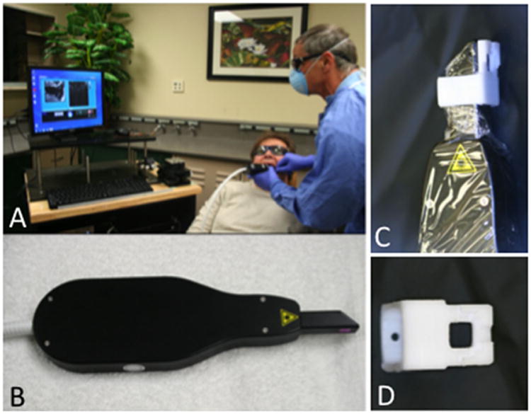
Introduction: Studies have shown that cross-polarization optical coherence tomography (CP-OCT) can be used to image the internal structure of carious lesions in vivo. The objective of this study was to show that CP-OCT can be used to monitor changes in the internal structure of early active carious lesions on smooth surfaces during non-surgical intervention with fluoride.
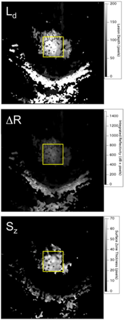
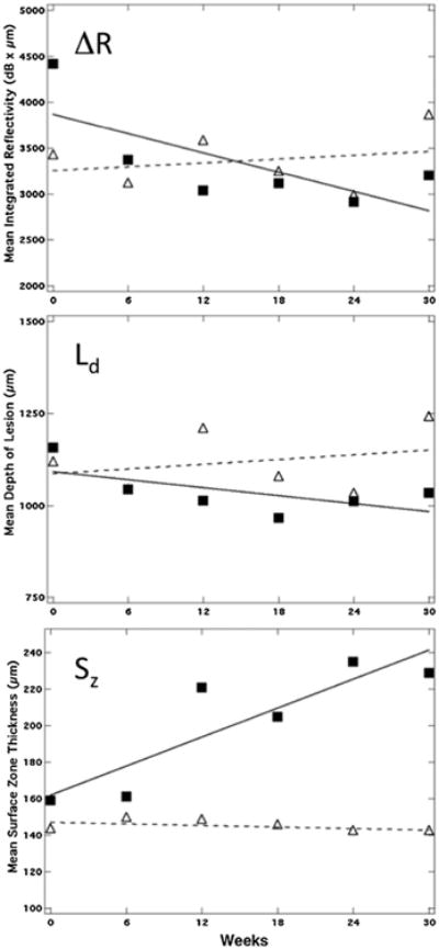
Methods: Lesions on the smooth surfaces of teeth were imaged using CP-OCT on 17 test subjects. Lesion structural changes were monitored during fluoride varnish application at 6-week intervals for 30 weeks. The lesion depth (Ld ), integrated reflectivity (ΔR), and surface zone thickness (Sz ) were monitored.
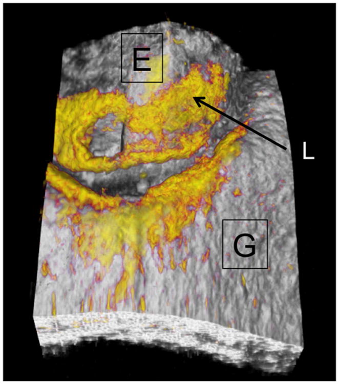
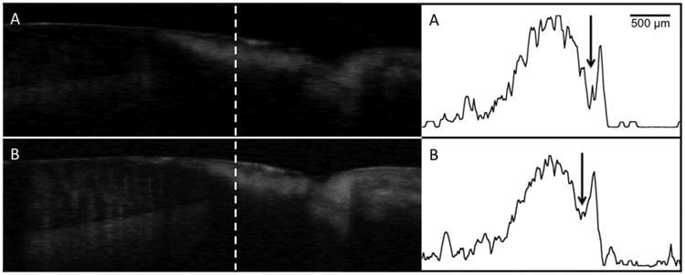
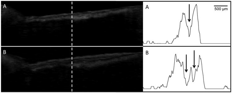
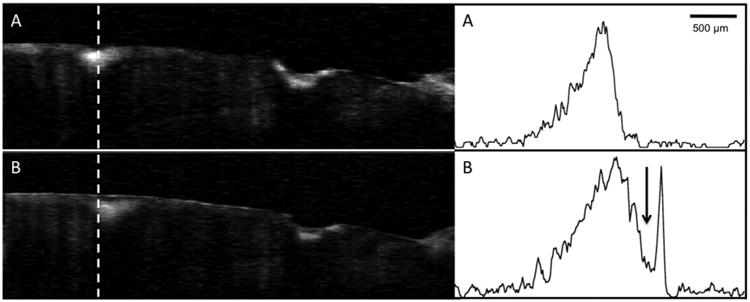
Results: A distinct transparent surface zone that may be indicative of lesion arrestment was visible in CP-OCT images on 62/63 lesions before application of fluoride varnish. The lesion depth and internal structure were resolved for all the lesions. The overall change in the mean values for Ld , ΔR, and Sz for all the lesions was minimal and was not significant during the study (P > 0.05). Only 5/63 lesions manifested a significant increase in Sz during intervention.
Conclusion: Even though it appears that most of the lesions manifested little change with fluoride varnish application in the 30 weeks of the study, CP-OCT was able to measure the depth and internal structure of all the lesions including the thickness of the important transparent surface zone located at the surface of the lesions, indicating that CP-OCT is ideally suited for monitoring lesion severity in vivo. Lasers Surg. Med. 48:915-923, 2016. © 2016 Wiley Periodicals, Inc.
Keywords: cross polarization optical coherence tomography; dental caries; tooth demineralization.
© 2016 Wiley Periodicals, Inc.
Conflict of interest statement
Conflict of Interest Disclosures: All authors have completed and submitted the ICMJE Form for Disclosure of Potential Conflicts of Interest and none were reported.
Figures
References
-
- Darling CL, Huynh GD, Fried D. Light scattering properties of natural and artificially demineralized dental enamel at 1310-nm. J Biomed Optics. 2006;11(3):034023 034021–034023 034011. - PubMed
-
- Fejerskov O, Kidd E. Dental caries: The disease and its clinical management. Oxford: Blackwell; 2003.
-
- Nyvad B, Machiulskiene V, Baelum V. Reliability of a new caries diagnostic system differentiating between active and inactive caries lesions. Caries Res. 1999;33(4):252–260. - PubMed
-
- Ismail A, Banting D, Eggertsson H, Ekstrand K, Ferreira-Zandona A, Longbottom C, Pitts N, Reich E, Ricketts D, Selwitz R, Sohn W, Topping G, Zero D. Rationale and evidence for the International Caries Detection and Assessment System (ICDAS II) proceedings of the 7th Indiana conference. 2005:161–221.
-
- Ekstrand KR, Ricketts DN, Longbottom C, Pitts NB. Visual and tactile assessment of arrested initial enamel carious lesions: An in vivo pilot study. Caries Res. 2005;39(3):173–177. - PubMed
Publication types
MeSH terms
Substances
Grants and funding
LinkOut - more resources
Full Text Sources
Other Literature Sources
Medical
Research Materials
Miscellaneous
