Review
. 2016 May;17(5):267-79.
doi: 10.1038/nrm.2016.7. Epub 2016 Mar 9.

Molecular circuitry of stem cell fate in skeletal muscle regeneration, ageing and disease

Affiliations
Review

Molecular circuitry of stem cell fate in skeletal muscle regeneration, ageing and disease

Albert E Almada et al. Nat Rev Mol Cell Biol. 2016 May.

Abstract

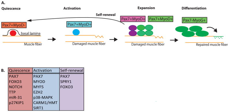
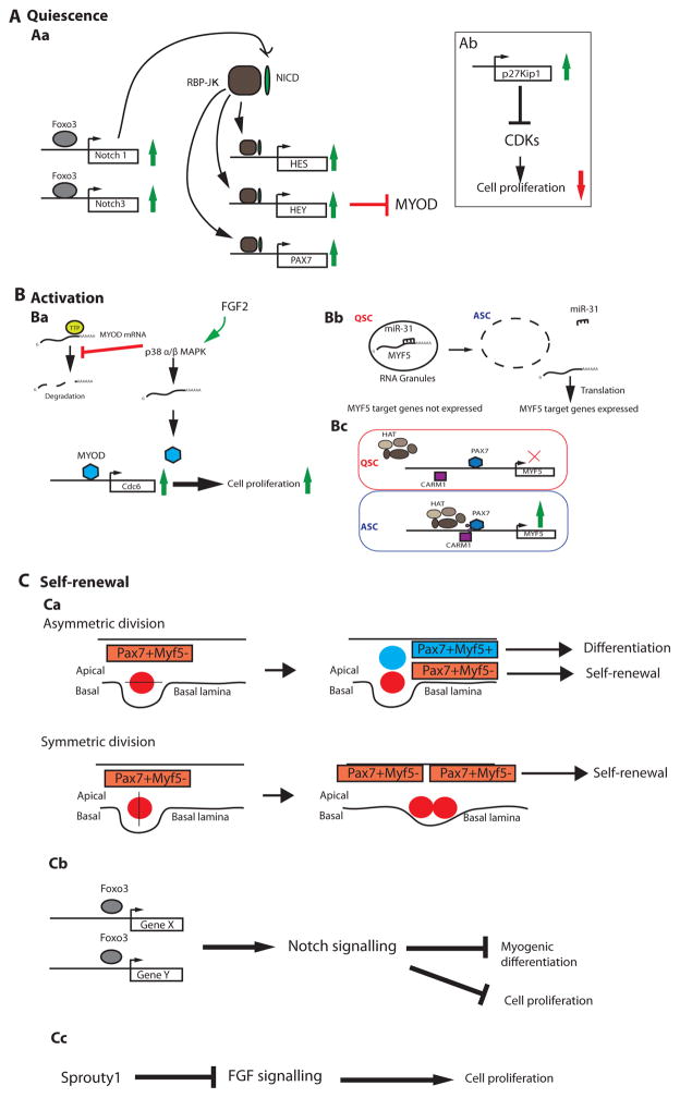
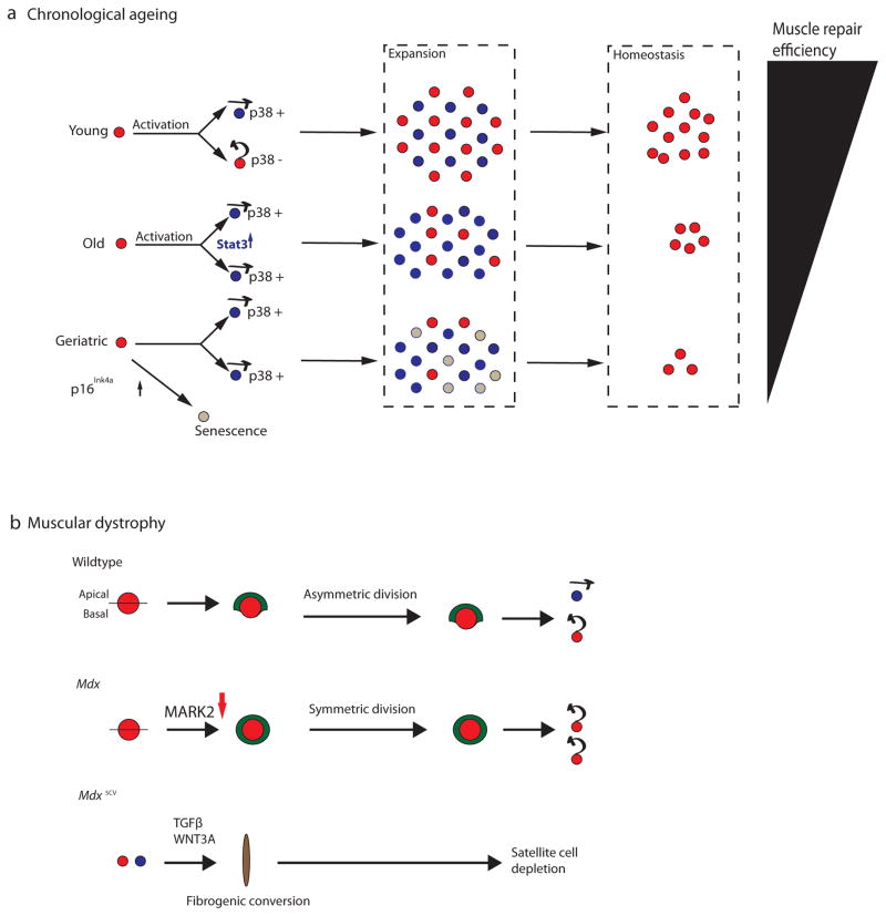
Satellite cells are adult myogenic stem cells that repair damaged muscle. The enduring capacity for muscle regeneration requires efficient satellite cell expansion after injury, their differentiation to produce myoblasts that can reconstitute damaged fibres and their self-renewal to replenish the muscle stem cell pool for subsequent rounds of injury and repair. Emerging studies indicate that misregulation of satellite cell fate and function can contribute to age-associated muscle dysfunction and influence the severity of muscle diseases, including Duchenne muscular dystrophy (DMD). It has also become apparent that satellite cell fate during muscle regeneration and ageing, and in the context of DMD, is governed by an intricate network of intrinsic and extrinsic regulators. Targeted manipulation of this network may offer unique opportunities for muscle regenerative medicine.


Figures

Figure 1
Figure 1. Classical view of muscle myogenesis
(A) During muscle homeostasis, satellite cells are maintained in a quiescent state (red) beneath the basal lamina and above the muscle sarcolemma of the muscle fiber. Quiescent satellite cells express Pax7 and lack MyoD. Upon muscle insult (ruptured fiber), satellite cells become activated (blue) and proliferate. Proliferating satellite cells maintain expression of Pax7 and induced expression of MyoD. A subset of these dividing satellite cells commit to differentiation (green), through the expression of Myogenin (MyoG) and downregulation of Pax7, ultimately producing myoblasts that exit the cell cycle and fuse to repair muscle. Alternatively, another subset of activated satellite cells will self-renew (purple) by inhibiting myogenic determination and re-instating quiescence. This ensures that the muscle retains a sufficient number of functional muscle stem cells to support future rounds of muscle repair. (B) Cell-intrinsic factors discussed in this review having roles in satellite cell quiescence, activation, and self-renewal.
Figure 2
Figure 2. Emerging regulators of satellite cell quiescence, activation, and self-renewal
(A)During homeostasis most satellite cells are quiescent. (Aa) The Notch signalling pathway plays a key role in enforcing satellite cell quiescence. Upon ligand/receptor binding, the Notch Intracellular Domain (NICD, pink oval) undergoes several proteolytic cleavages (not shown), and then translocates to the nucleus where it interacts with the Recombining Binding Protein J-κ effector protein (RBPJ-κ, blue circle). The NICD:RBPJ-κ complex activates expression of target genes, including the Hes and Hey family of transcription factors, which function to inhibit MYOD. RBPJ-κ also binds the promoter of Pax7 and induces its expression. PAX7 induction further downregulates MYOD via transcription-independent mechanisms (not shown). The Forkhead box 3 (FOXO3, green circle) transcription factor can induce expression of NOTCH1 and NOTCH3 trans-membrane proteins, which facilitates Notch signalling by ensuring an adequate number of receptors are produced and shuttled to the cell membrane where they can interact with available ligands in the local environment. (Ab) Satellite cell entry into the cell cycle is inhibited through the activities of p27Kip1, which inhibits cyclin-dependant kinases (CDKs). (B) After muscle injury, activated satellite cells detect pro-myogenic stimuli from the local environment and begin proliferation. (Ba) Exposure to FGF2 induces p38α/β mitogen-activated protein kinase (MAPK) activity, which inhibits the RNA-binding destabilizing protein, Tristetraprolin (TTP), leading to stabilization of the MyoD mRNA. This allows MYOD protein to accumulate (purple circle) and induce expression of Cdc6, a DNA replication gene that promotes cell cycle entry and thus proliferation. (Bb) Another activation regulator, MYF5, is post-transcriptionally repressed by miRNA-31 (miR-31), which sequesters Myf5 mRNA into RNA granules in quiescent satellite cells (QSCs) and prevents activation of MYF5 targets. Upon muscle trauma, the RNA granules dissociate, miRNA-31 levels decrease, and MYF5 protein is produced and can activate target genes. (Bc) The Myf5 gene is also transcriptionally regulated through the activities of CARM1 (pink circle), an arginine methyl-transferase that methylates PAX7 (small purple circles on the light blue circle) leading to PAX7 association with the Histone Acetyl-Transferase complex (HAT, purple complex), and the induced expression of target genes (such as MYF5) in activated satellite cells (ASCs). (C) Satellite cells are capable of self renewal. (Ca) A balance between self-renewal and differentiation of satellite cells is acheived by the mode of cell division. Apico-basal divisions generate one self-renewing (red, Myf5-) and one differentiating cell (blue, Myf5+). On the other hand, planar divisions (parallel to the muscle fiber) are symmetric and generate two daughters that retain self-renewal capabilities. (Cb) Self-renewal of satellite cells can be followed by their return to quiescence. FOXO3 induces expression of Notch signalling genes, enabling the cascade of molecular events leading to the inhibition of MYOD-mediated differentiation and blockade of cell cycle progression. (Cc) The return to quiescence also requires Sprouty1 (Spry1), a tyrosine kinase inhibitor that inhibits FGF signalling, thereby restricting cellular proliferation.
Figure 3
Figure 3. Intrinsic cell fate regulators are misregulated in aged and dystrophic satellite cells
(A) In youth and adulthood (age 2–6 months in mice), quiescent satellite cells (red circles) asymmetrically distribute p38 to daughter cells, such that one daughter receives p38 and is competent for myogenic commitment and differentiation (blue circle with arrow pointing forwards to denote lineage commitment), while the other daughter self-renews and returns to the quiescent state (red circle with arrow pointing backwards to denote their self-renewal and return to quiescence). Asymmetric satellite cell divisions in young animals yield a healthy balance between satellite cells that are competent for myogenic differentiation and cells that self-renew during the expansion phase, which ensures the efficient repair of muscle and the return of a sufficient number of cells to the satellite cell niche to support future demands for muscle regeneration (homeostasis). However, in aged animals (18–24 months in mice), p38 is symmetrically partitioned to daughter cells and JAK-STAT signalling is elevated in proliferating cells, leading to an overall increase in myogenic commitment within the satellite cell population, and reducing the number of quiescent satellite cells at homeostasis. In “geriatric” muscle (28–32 months of age in mice), p16Ink4a is additionally elevated in the satellite cell population, leading to a block in satellite cell activation and directing these cells to an irreversible state of cellular senescence (grey circles). The intrinsic deficits in aged and “geriatric” satellite cells deplete the self-renewing satellite cell population at homeostasis, and thus impair the regenerative response in skeletal muscle as animals age (Black triangle). (B) Satellite cells from 2-month old animals (adult) carrying a loss-of-function mutation in the Dystrophin gene (mdx) display defects in asymmetric division and a stark reduction in apico-basally oriented divisions due to reduced levels of polarity kinase MARK2 (also known as Par-1). This alters the localization of PARD3 (shown in green) and impairs mitosis, leading to a failure to properly expand the lineage committed satellite cells, and a consequent inefficiency of muscle repair in response to injury. In mice carrying an alternative mdx allele (mdx5cv ), dystrophic satellite cells (either QSC or ASC) can undergo a cell type transition towards a fibrogenic state (brown oval) via overactivation of Wnt and TGFβ signalling. This may over time and in a context-dependant manner lead to depletion of highly functional muscle stem cells.

References

    1. Mauro A. Satellite cell of skeletal muscle fibers. J Biophys Biochem Cytol. 1961;9:493–5. The first reported observation of the satellite cell in skeletal muscle in frog. - PMC - PubMed
    1. Bentzinger CF, et al. Fibronectin regulates Wnt7a signaling and satellite cell expansion. Cell Stem Cell. 2013;12:75–87. - PMC - PubMed
    1. Yin H, Price F, Rudnicki MA. Satellite cells and the muscle stem cell niche. Physiol Rev. 2013;93:23–67. - PMC - PubMed
    1. Urciuolo A, et al. Collagen VI regulates satellite cell self-renewal and muscle regeneration. Nat Commun. 2013;4:1964. - PMC - PubMed
    1. Brohl D, et al. Colonization of the satellite cell niche by skeletal muscle progenitor cells depends on Notch signals. Dev Cell. 2012;23:469–81. - PubMed

Substances