Review
. 2016 May;17(5):308-21.
doi: 10.1038/nrm.2016.14. Epub 2016 Mar 9.

Nuclear DNA damage signalling to mitochondria in ageing

Affiliations
Review

Nuclear DNA damage signalling to mitochondria in ageing

Evandro Fei Fang et al. Nat Rev Mol Cell Biol. 2016 May.

Abstract

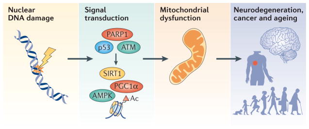
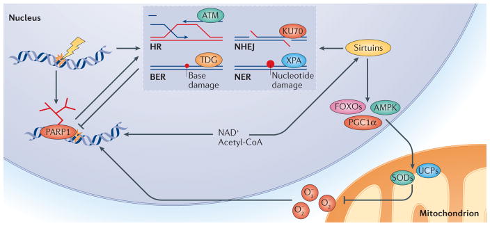
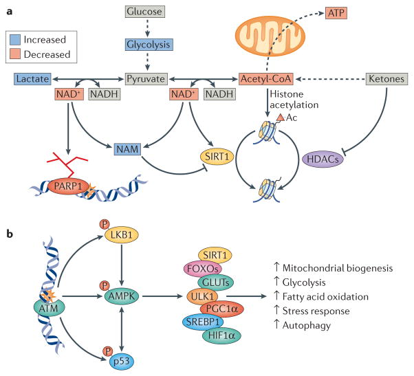
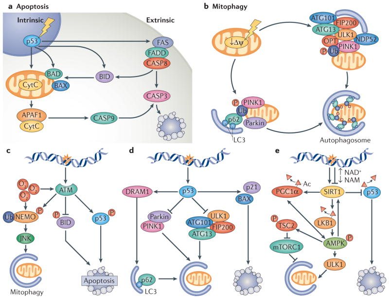
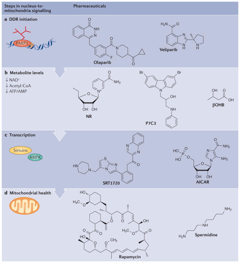
Mitochondrial dysfunction is a hallmark of ageing, and mitochondrial maintenance may lead to increased healthspan. Emerging evidence suggests a crucial role for signalling from the nucleus to mitochondria (NM signalling) in regulating mitochondrial function and ageing. An important initiator of NM signalling is nuclear DNA damage, which accumulates with age and may contribute to the development of age-associated diseases. DNA damage-dependent NM signalling constitutes a network that includes nuclear sirtuins and controls genomic stability and mitochondrial integrity. Pharmacological modulation of NM signalling is a promising novel approach for the prevention and treatment of age-associated diseases.


Conflict of interest statement

statement The authors declare competing interests: see Web version for details.

Figures

Figure 1
Figure 1. An overview of DNA damage-induced nucleus-to-mitochondria signalling and ageing
Nuclear DNA damage can cause mitochondrial dysfunction. DNA damage leads to the activation of a number of proteins, resulting in widespread downstream changes in various cellular pathways. Among these pathways, signalling from the nucleus to mitochondria (NM signalling) is less studied than many others, but it may be very important in the ageing process and in age-associated diseases, and interventions in this process may slow ageing. A number of factors contribute to NM signalling, such as upstream DNA damage sensors including poly(ADP-ribose) polymerase 1 (PARP1), ataxia telangiectasia mutated (ATM) and the transcription factor p53. The NAD-dependent protein deacetylase sirtuin 1 (SIRT1) and the transcription regulator AMP-activated protein kinase (AMPK) then propagate the NM signal through post-translational modifications (deacetylation or phosphorylation, respectively) of DNA histones, peroxisome proliferator-activated receptor-γ co-activator 1α (PGC1α) and other proteins. The combined effect of the activation of the DNA damage response is downstream changes in the transcriptome, epigenome and metabolome, and in cellular bioenergetics. These contribute to ageing and the development of age-associated diseases such as neurodegeneration and cancer.
Figure 2
Figure 2. PARP1–NAD+–SIRT1-mediated nuclear DNA damage to mitochondria signalling
Activation of poly(ADP-ribose) polymerase 1 (PARP1) upon DNA damage facilitates DNA repair but leads to loss of NAD+ and acetyl-CoA, the latter being an important molecule in cellular metabolism. This results in inhibition of sirtuins such as SIRT1, owing to competition for NAD+. Loss of sirtuin activity leads to an increase in mitochondrial reactive oxygen species (O2), owing to decreased activation of downstream stress response factors such as AMP-activated protein kinase (AMPK), forkhead box O proteins (FOXOs) and peroxisome proliferator-activated receptor-γ co-activator 1α (PGC1α). Collectively, these enzymes regulate a large number of genes that are involved in coping with oxidative stress, such as those encoding uncoupling proteins (UCPs) like UCP2 and superoxide dismutases (SODs). In addition, SIRT1, as well as SIRT6, has been shown to positively regulate several DNA repair pathways, such as homologous recombination (HR), non-homologous end-joining (NHEJ), base excision repair (BER) and nucleotide excision repair (NER). Specifically, SIRT1 has been shown to deacetylate repair factors such as xeroderma pigmentosum group A-complementing protein (XPA, which is involved in NER), KU70 (involved in NHEJ), ataxia telangiectasia mutated (ATM; involved in HR and NHEJ) and thymine DNA glycosylase (TDG; involved in BER). Activation of PARP1 and subsequently less-robust DNA repair, as well as increased oxidative stress, may represent a vicious cycle that could contribute to the ageing process. Thus, PARP1 is pivotal to the initiation of different DNA repair pathways, and it interacts with SIRT1 to execute cellular signalling from the nucleus to mitochondria.
Figure 3
Figure 3. DNA damage-induced nucleus-to-mitochondria signalling is linked to metabolic dysfunction and age-associated diseases
a | Poly(ADP-ribose) polymerase 1 (PARP1) activation leads to alterations in central bioenergetic pathways. A consequence of loss of NAD+ is change in the NAD+/NADH ratio. Notably, PARP1 activation also leads to increased nicotinamide (NAM), a competitive inhibitor of sirtuins such as SIRT1, as well as loss of NAD+, which is a substrate for sirtuins. The decrease in the NAD+/NADH ratio also leads to the conversion of pyruvate to lactate by lactate dehydrogenase, while decreasing the formation of acetyl-CoA by pyruvate dehydrogenase. An isoform of pyruvate dehydrogenase has recently been found in the cell nucleus, and the DNA damage response could thereby regulate the formation of acetyl-CoA locally. Loss of acetyl-CoA decreases histone acetylation (Ac) and changes the epigenome, resulting in chromatin condensation and gene silencing, while also leading to decreased availability of acetyl-CoA for the mitochondria for the generation of ATP. Histone acetylation is also reduced by the activity of sirtuins such as SIRT1 and of other classes of histone Lys deacetylases (HDACs). Ketones, which are a group of metabolites made in the liver, increase acetyl-CoA levels and histone acetylation and could counteract the effects of age-associated DNA damage. b | Ataxia telangiectasia mutated (ATM) regulates multiple metabolic pathways following DNA damage through phosphorylation of p53, liver kinase B1 (LKB1) and AMP-activated kinase (AMPK). AMPK is a central kinase in the adaptive cellular response and phosphorylates a number of key factors. These include SIRT1, which is a known regulator of mitochondrial biogenesis; forkhead box O transcription factors (FOXOs), which are involved in cellular stress responses; glucose transporters (GLUTs), which are involved in glucose transport and glycolysis; UNC51-like kinase 1 (ULK1), which is a regulator of autophagy; peroxisome proliferator-activated receptor-γ co-activator 1α (PGC1α), which is a master-regulator of mitochondrial biogenesis; sterol regulatory element-binding protein 1 (SREBP1), which is a regulator of fatty acid oxidation; and hypoxia inducible factor 1α (HIF1α), which is a central positive regulator of glycolysis. Collectively the activity of these factors leads to changes in cellular bioenergetics.
Figure 4
Figure 4. DNA damage signalling in the regulation of mitophagy and apoptosis
a | Apoptosis can be initiated through intrinsic or extrinsic pathways. The intrinsic pathways can be initiated by genotoxic stress, leading to the activation of p53 and transcriptional upregulation of pro-apoptotic factors such as BAD, BAX and BID. If the stress is sustained or high enough, BAD and BAX accumulation results in the permeabilization of the outer mitochondrial membrane and release of cytochrome C (CytC) that can associate with apoptosis-activating factor 1 (APAF1) and activate caspase 9 (CASP9). CASP9 activation leads to CASP3 activation and initiation of apoptosis. The extrinsic pathway can be initiated through activation of the FAS receptor. This leads to FAS-associated death domain protein (FADD)-mediated activation of CASP8, resulting in CASP3 activation. Notably, there is considerable crosstalk between intrinsic and extrinsic pathways. b | Selective mitophagy is regulated through factors such as PTEN-induced putative kinase 1 (PINK1) and the E3-ubiquitin ligase parkin. Initially, mitochondrial damage causes loss of the mitochondrial membrane potential (Δψ). This leads to the retention of PINK1 at the outer mitochondrial membrane and phosphorylation of outer membrane proteins. Concomitantly, parkin is activated, leading to ubiquitylation of outer membrane proteins. PINK1 phosphorylates ubiquitin that recruits adaptor proteins such as optineurin (OPT), nuclear dot protein 52 (NDP52) and p62, leading to recruitment of the UNC51-like kinase 1 (ULK1) complex (consisting of proteins such as autophagy-related protein 101 (ATG101), ATG13 and FIP200) and to the formation of a double lipid membrane-bound vesicle, the autophagosome. The adaptor proteins (OPT, p62 and NDP52) interact with the autophagosome through light chain 3 (LC3), a small protein that coats the autophagosome. The entire mitochondrion will eventually become engulfed in the autophagosome, which will fuse with a lysosome leading to degradation of the mitochondrion. c | Ataxia telangiectasia mutated (ATM) is activated by breaks in DNA and possibly also by oxidative stress (O2). At low levels of DNA damage stress, ATM activation leads to phosphorylation, ubiquitylation and activation of the NEMO JNK (NF-κB essential modulator Jun N-terminal kinase) pathway that stimulates mitophagy. ATM also phosphorylates the pro-apoptotic factor BID to inhibit apoptotic signalling. At high levels of stress, ATM phosphorylates and activates p53, which propagates pro-apoptotic signals. d | p53 has been well characterized in the response to genotoxic stimuli, in which it transcriptionally activates pro-apoptotic proteins such as BAX and p21 while simultaneously inhibiting the ULK1-containing autophagy-initiating complex. In addition, p53 activation may lead to decreased expression of parkin and decreased activation of parkin PINK1-mediated mitophagy. At lower levels of stress, p53 can stimulate mitophagy through the activation of DNA damage-regulated autophagy modulator protein 1 (DRAM1), which stimulates p62-mediated mitophagy. e | Sirtuin 1 (SIRT1) regulates both mitophagy and apoptosis. At low levels of DNA damage, nuclear SIRT1 can be activated to facilitate DNA repair. After lethal levels of nuclear DNA damage, SIRT1 is inhibited by the DNA damage response, leading to p53 acetylation and cell death. Loss of SIRT1 activity decreases the stimulation of mitophagy through peroxisome proliferator-activated receptor-γco-activator 1α (PGC1α)- and AMP-activated kinase (AMPK)-dependent pathways. SIRT1 regulates mitochondrial biogenesis and mitophagy through deacetylation of PGC1α, and it also interacts with AMPK to regulate mitophagy through mutual activation: AMPK activates SIRT1 by increasing the ratio of NAD+/NADH, and it is activated by SIRT1 through deacetylation of liver kinase B1 (LKB1), an AMPK activator. Furthermore, AMPK phosphorylates and activates the mTORC1 inhibitor protein tuberous sclerosis complex 2 (TSC2), thereby further activating autophagy. AMPK also positively regulates PGC1α activity, which stimulates mitochondrial biogenesis and activates forkhead box protein O 3A, leading to activation of stress response and pro-survival pathways. NAM, nicotinamide.
Figure 5
Figure 5. Pharmacological interventions in the DNA damage response that may lead to increases in lifespan and healthspan
a | Inhibition of poly(ADP-ribose) polymerase 1 (PARP1) by olaparib and other PARP inhibitors has been shown to increase lifespan in model organisms. b | Impaired cellular metabolism can be counteracted by treatment with NAD+ precursors such as nicotinamide riboside (NR), by activation of NAD+-generating enzymes such as nicotinamide phosphoribosyltransferase by P7C3, or by replenishment of acetyl-CoA levels with ketones such as β-hydroxybutyrate (βOHB). c | Signalling factors and targets in the DNA damage response (DDR) may constitute additional targets for interventions. These include activation of sirtuin 1 by compounds such as SRT1720, or AMP-activated kinase (AMPK) activation using the AMP analogue AICAR. d | Mitochondrial function may be augmented by stimulation of autophagy by rapamycin or its newer analogues, or by stimulation of autophagy through alternative pathways using compounds such as spermidine.

References

    1. West AP, et al. Mitochondrial DNA stress primes the antiviral innate immune response. Nature. 2015;520:553–557. - PMC - PubMed
    1. Wallace DC. Mitochondrial DNA variation in human radiation and disease. Cell. 2015;163:33–38. - PMC - PubMed
    1. Scheibye-Knudsen M, et al. Protecting the mitochondrial powerhouse. Trends Cell Biol. 2014;25:158–170. - PMC - PubMed
    1. Maynard S, Fang EF, Scheibye-Knudsen M, Croteau DL, Bohr VA. DNA damage, DNA repair, aging, and neurodegeneration. Cold Spring Harb Perspect Med. 2015;5:a025130. - PMC - PubMed
    1. Youle RJ, van der Bliek AM. Mitochondrial fission, fusion, and stress. Science. 2012;337:1062–1065. - PMC - PubMed

Substances