. 2016 Apr;5(4):451-63.
doi: 10.5966/sctm.2015-0155. Epub 2016 Mar 8.

Impaired Angiogenic Potential of Human Placental Mesenchymal Stromal Cells in Intrauterine Growth Restriction

Affiliations

Impaired Angiogenic Potential of Human Placental Mesenchymal Stromal Cells in Intrauterine Growth Restriction

Chiara Mandò et al. Stem Cells Transl Med. 2016 Apr.

Abstract

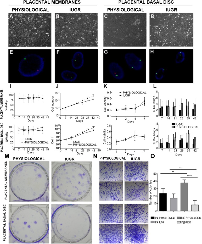
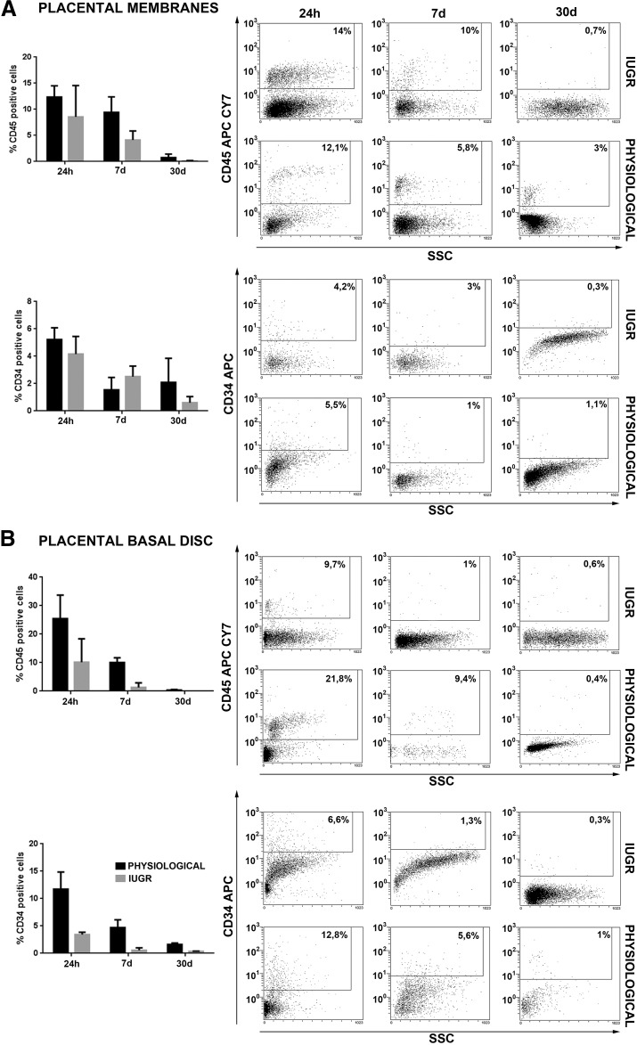
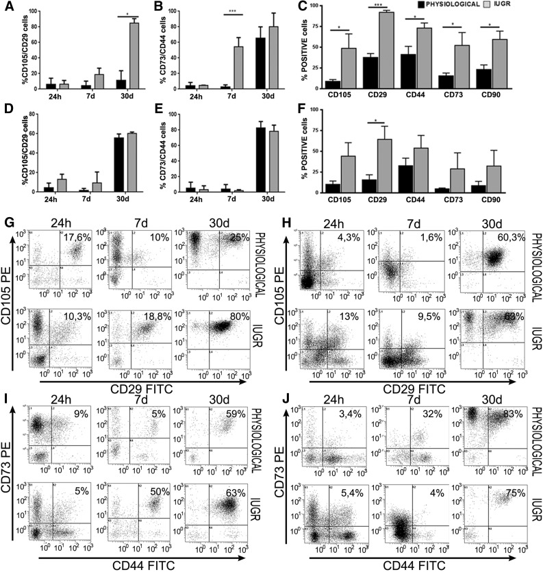
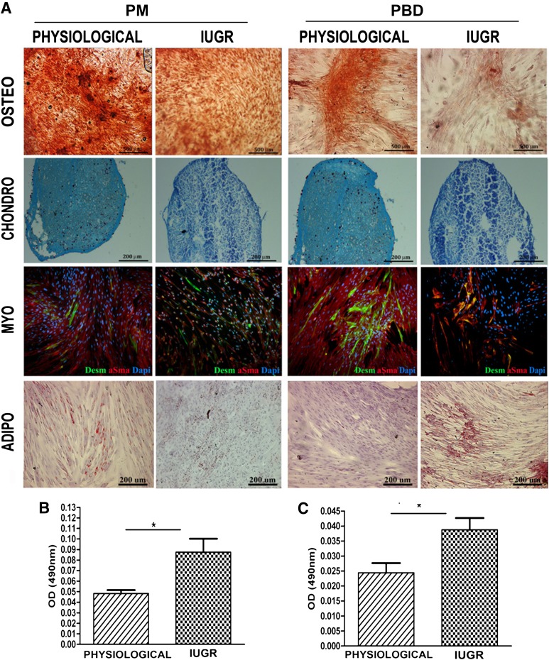
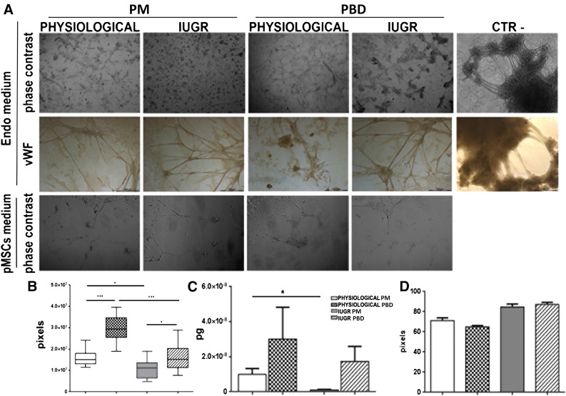
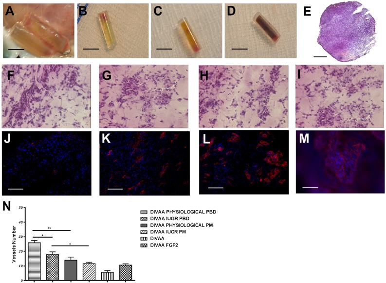
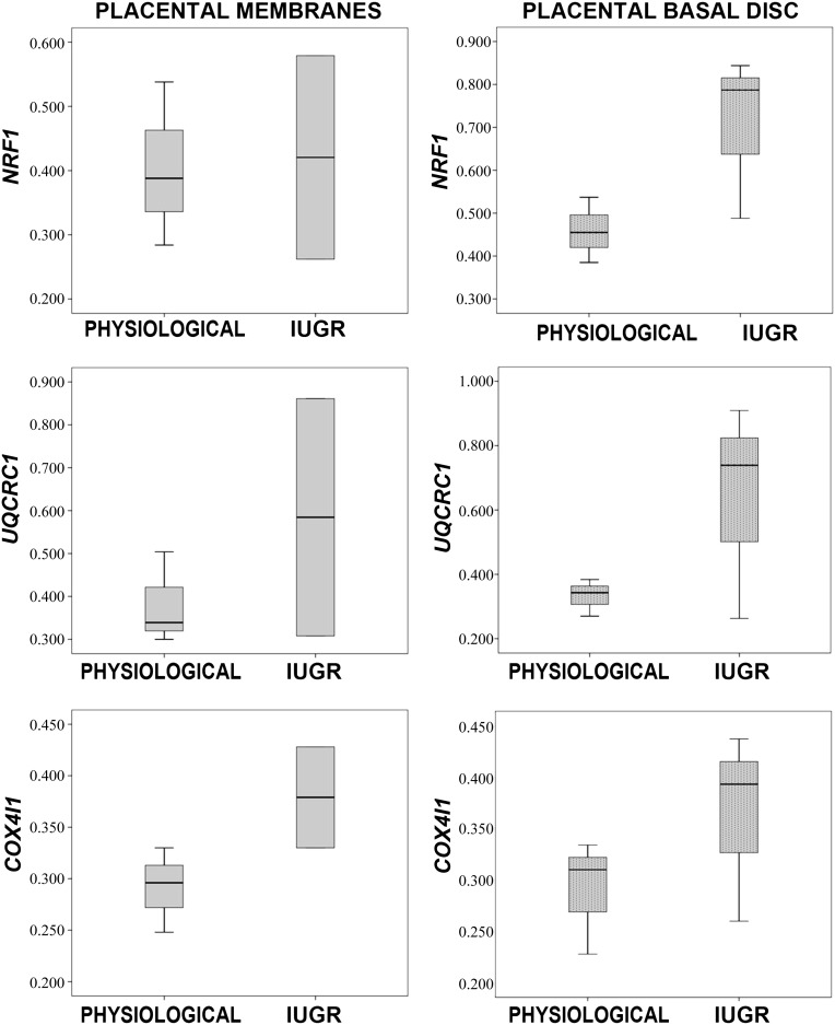
Human placental mesenchymal stromal cells (pMSCs) have never been investigated in intrauterine growth restriction (IUGR). We characterized cells isolated from placental membranes and the basal disc of six IUGR and five physiological placentas. Cell viability and proliferation were assessed every 7 days during a 6-week culture. Expression of hematopoietic, stem, endothelial, and mesenchymal markers was evaluated by flow cytometry. We characterized the multipotency of pMSCs and the expression of genes involved in mitochondrial content and function. Cell viability was high in all samples, and proliferation rate was lower in IUGR compared with control cells. All samples presented a starting heterogeneous population, shifting during culture toward homogeneity for mesenchymal markers and occurring earlier in IUGR than in controls. In vitro multipotency of IUGR-derived pMSCs was restricted because their capacity for adipocyte differentiation was increased, whereas their ability to differentiate toward endothelial cell lineage was decreased. Mitochondrial content and function were higher in IUGR pMSCs than controls, possibly indicating a shift from anaerobic to aerobic metabolism, with the loss of the metabolic characteristics that are typical of undifferentiated multipotent cells.

Significance: This study demonstrates that the loss of endothelial differentiation potential and the increase of adipogenic ability are likely to play a significant role in the vicious cycle of abnormal placental development in intrauterine growth restriction (IUGR). This is the first observation of a potential role for placental mesenchymal stromal cells in intrauterine growth restriction, thus leading to new perspectives for the treatment of IUGR.

Keywords: Intrauterine growth restriction; Mesenchymal stromal cells; Placenta; Pregnancy; Stem cells.


Figures

Figure 1.
Figure 1.
Characterization and proliferation of placental mesenchymal stromal cells (pMSCs). (A–D): Morphology of pMSCs at passage 3 isolated from the indicated placental compartment (physiological, n = 5; IUGR n = 6) observed by phase-contrast microscopy (original magnification, ×10). Fluorescent in situ hybridization analysis shows a male pattern (E, F), a mixed male/female pattern (G), and a female pattern (H). (I): Viability of cells isolated from physiological (n = 5 each group) and IUGR (n = 6 each group) PM and PBD placentas after 6 weeks of culture in proliferation medium. DXZ1 (Xp11.1q11.1) spectrum green; SRY (Yp11.3) spectrum orange. (J): Expansion rate of the same samples observed during 6 weeks of culture obtained by visual counting after trypan blue staining. Data are shown as average ± SD (in percentages) of 11 placental samples, each performed in three replicates. ∗, p < .01; ∗∗∗, p < .001. (K): Proliferative potential of pMSCs isolated from physiological and IUGR PM and PBD cultured at passage 3 (P3) was measured by 3-[31]-2,5-diphenyltetrazolium bromide assay after 0, 1, 2, 4, and 7 days of seeding. (L): Measurements of doubling rate every 7 days, during the entire culture period, based on visual count after trypan blue staining. (M): Crystal violet-stained plates of colony-forming units-fibroblast (CFU-F) assays performed on P3 pMSCs isolated from physiological and IUGR PM and PBD. (N): Representative phase-contrast microscopic images of single CFU-F assay of P3 pMSCs from physiological and IUGR PM and PBD stained by crystal violet (original magnification, ×4). (O): CFU-F assay analysis. The data represent the mean ± SEM number of colonies per plate. Abbreviations: IUGR, intrauterine growth restriction; PBD, placental basal disc; PM, placental membrane.
Figure 2.
Figure 2.
Flow cytometry analysis for hematopoietic antigen expression in placental mesenchymal stromal cells (pMSCs). FACScan (BD Biosciences, Franklin Lakes, NJ, http://www.bdbiosciences.com) immunophenotyping of human placental membrane- and placental basal disc (PBD)-derived cells of physiological (n = 5 each group) and IUGR (n = 6 each group) placentas. Histograms show representative examples of placental cells isolated stained with cell surface markers. Hematopoietic markers expression was analyzed 24 hours after isolation and after 7 and 30 days of culture (A, B). pMSCs expressed hematopoietic markers after 24 hours of culture, which decreased considerably after 7 and 30 days of culture. The reduction of CD34 and CD45 hematopoietic markers expression was more evident in pMSC isolated from PBD-derived cells of physiological and IUGR placentas (B). Abbreviations: APC, allophycocyanin; IUGR, intrauterine growth restriction; SSC, side scatter.
Figure 3.
Figure 3.
Flow cytometry analysis for mesenchymal antigen expression in placental mesenchymal stromal cells (pMSCs). FACScan (BD Biosciences) immunophenotyping of human placental membrane (PM)- and placental basal disc (PBD)-derived cells of physiological (n = 5 each group) and intrauterine growth restriction (IUGR) (n = 6 each group) placentas. (A, B, D, E): Histograms show representative examples of pMSCs isolated from passage 3 stained with mesenchymal cell surface markers. Coexpression of CD105/CD29 and CD44/CD73 increased during PM and PBD cell culture. At day 7, mesenchymal markers were 1.6–4-fold (membranes) and 2.3–5.8-fold (basal disc) higher in IUGR placentas compared with controls. Differences were statistically significant for CD105, CD44, CD73, and CD90 (p < .05) and for CD29 (p < .001) in placental membranes (C) and only for CD29 (p < .05) in placental basal disc (F). ∗, p < .05; ∗∗∗, p < .001. Moreover, dot plots show representative examples of coexpression of more significant mesenchymal markers, such as CD105+ CD29+ and CD44+CD73+ at different days of culture (G–J). After 30 days, all samples showed a typical homogeneous immunophenotype of mesenchymal stromal cells. Abbreviations: FITC, fluorescein isothiocyanate; PE, preeclampsia.
Figure 4.
Figure 4.
Placental mesenchymal stromal cell (pMSC) multilineage differentiation in vitro: osteogenic, chondrogenic, adipogenic, and myogenic commitment. pMSC isolated from placenta membrane and placental basal disc physiological (n = 5 each group) and IUGR (n = 6 each group) placentas at passage 3 were investigated for their in vitro multilineage differentiation capacity. (A): Osteogenesis was demonstrated by enhancement of the alizarin red staining. Chondrogenesis was indicated by alcian blue staining in cryosections from pMSCs. Desmin (green) and α-smooth muscle acti (red) staining confirmed their myogenic differentiation. Nuclei were stained blue with 4′,6-diamidino-2-phenylindole. Adipogenesis was detected by the formation of neutral lipid vacuoles stainable with Oil-Red O (for all representative examples, original magnification, ×10). (B, C): Adipogenic differentiation was quantified by spectrophotometric measurements with Oil Red O levels. ∗, p < .05. Abbreviations: adipo, adipogenic; chondro, chondrogenic; Dapi, 4′,6-diamidino-2-phenylindole; IUGR, intrauterine growth restriction; myo, myogenic; osteo, osteogenic.
Figure 5.
Figure 5.
Tubulo formation assay. Endothelial differentiation potential of pMSCs was analyzed by immunofluorescent and immunohistochemistry assays. (A): Phase-contrast morphology of PM-derived pMSCs isolated from physiological (n = 5) and IUGR (n = 6) and PBD-derived pMSCs isolated from physiological (n = 5) and IUGR (n = 6) and human fibroblasts used as negative control after 72 hours of two-dimensional endothelial differentiation (original magnification, ×10). Physiological pMSCs formed more capillary-like tube structures than IUGR pMSCs. In fibroblast cells, we never observed tubular structures. These data were confirmed by quantification of endothelial differentiation, measuring tube-like structure length (B), histological coloration with vWF (A), and CD31 expression by real-time polymerase chain reaction analysis (C). ∗, p < .05; ∗∗, p < .005; ∗∗∗, p < .001. The angiogenic effect of pMSCs was indirectly measured by incubating pMSC-conditioned medium with human umbilical vein endothelial cell line in a Matrigel tube formation assay (A) and quantified by measuring tube-like structure length (D). Abbreviations: IUGR, intrauterine growth restriction; CTR-, negative control; Endo medium, two-dimensional endothelial differentiation; PD, placental basal disc; PM, placental membrane; pMSCs medium, placental mesenchymal stromal cell-conditioned medium; vWF, von Willebrand factor.
Figure 6.
Figure 6.
Angiogenic potential of placental mesenchymal stromal cells (pMSCs) isolated from human physiological and intrauterine growth restriction (IUGR) placentas. Quantification of angiogenic activity of PM- and PBD-derived pMSCs isolated from physiological and IUGR performed by the DIVAA system. (A): Angioreactors were recovered at 15 days after subcutaneous implantation. Unlike the control angioreactors with no angiogenic factor (B), assays containing an angiogenic factor (fibroblast growth factor-2 [FGF-2]) (C) and human pMSCs only (D) revealed numerous blood vessels near the open end that invade the Matrigel. Sections of paraffin-embedded angioreactor containing pMSCs stained by hematoxylin and eosin (H&E) (E) (original magnification, ×10), and H&E staining of angioreactor containing physiological and IUGR PBD-derived pMSCs (F, G) and physiological and IUGR PM-derived pMSCs (H, I) (original magnification, ×40). Matrigel invasion and early organization of endothelial cell-containing structures is revealed by vWF (in red) (angioreactor only [J], angioreactor plus FGF-2 [K], and the angioreactor with physiological and IUGR PBD-derived pMSCs [L, M]). PBD-derived pMSCs isolated from physiological and IUGR spread throughout the surface and aligned to form branching, anatomizing tubes with multicentric junctions that formed a closely knit meshwork of capillary-like structures (L, M). The number of von Willebrand factor-positive vessels represented in (N) was obtained by counting six fields at original magnification, ×20 (∗, p < .05; ∗∗, p < .005). Abbreviations: DIVAA, directed in vivo angiogenesis angioreactor; PD, placental basal disc; PM, placental membrane.
Figure 7.
Figure 7.
Gene expression analysis of mitochondrial biogenesis activator and respiratory chain complexes of placental mesenchymal stromal cells (pMSCs). Gene expression of NRF1 (mitochondrial biogenesis activator), UQCRC1, and COX4I1 (mitochondrial respiratory chain subunits) in placental membrane- and placental basal disk-derived pMSCs isolated from five physiological and six IUGR pregnancies. Relative expression values are shown as box plots, indicating the median and the 25th and 75th percentiles. Abbreviation: IUGR, intrauterine growth restriction.

Similar articles

Cited by

References

    1. Nuzzo AM, Giuffrida D, Zenerino C, et al. JunB/cyclin-D1 imbalance in placental mesenchymal stromal cells derived from preeclamptic pregnancies with fetal-placental compromise. Placenta. 2014;35:483–490. - PubMed
    1. Dominici M, Le Blanc K, Mueller I, et al. Minimal criteria for defining multipotent mesenchymal stromal cells. The International Society for Cellular Therapy position statement. Cytotherapy. 2006;8:315–317. - PubMed
    1. Brooke G, Tong H, Levesque JP, et al. Molecular trafficking mechanisms of multipotent mesenchymal stem cells derived from human bone marrow and placenta. Stem Cells Dev. 2008;17:929–940. - PubMed
    1. Yen BL, Huang HI, Chien CC, et al. Isolation of multipotent cells from human term placenta. Stem Cells. 2005;23:3–9. - PubMed
    1. Abumaree MH, Al Jumah MA, Kalionis B, et al. Phenotypic and functional characterization of mesenchymal stem cells from chorionic villi of human term placenta. Stem Cell Rev. 2013;9:16–31. - PubMed

Publication types