Case Reports
. 2016 Oct;24(10):1511-4.
doi: 10.1038/ejhg.2016.22. Epub 2016 Mar 9.

Somatic mosaicism due to a reversion variant causing hemi-atrophy: a novel variant of dystrophinopathy

Affiliations
Case Reports

Somatic mosaicism due to a reversion variant causing hemi-atrophy: a novel variant of dystrophinopathy

Jaya Punetha et al. Eur J Hum Genet. 2016 Oct.

Abstract

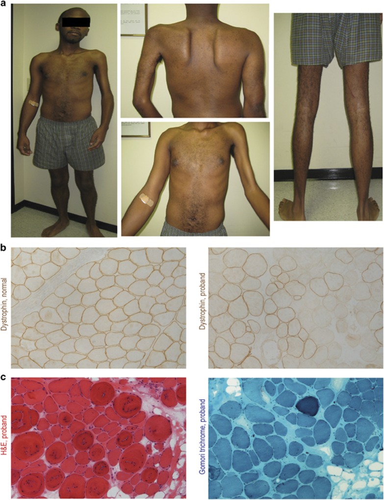
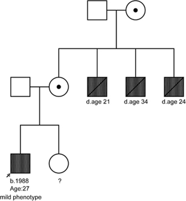
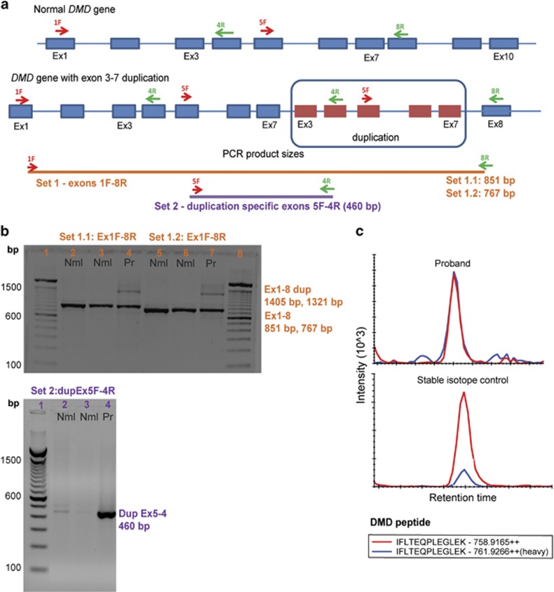
We describe a case of hemi-atrophy in a young adult male, with a positive family history of three maternal uncles with Duchenne muscular dystrophy (DMD). The patient showed progressive weakness localized to the left side, an abnormal electromyography, and creatine kinase levels >3000 IU/l. Muscle biopsy showed both dystrophin-positive and -negative myofibers. An out-of-frame duplication variant in DMD, that is, c.(93+1_94-1)_(649+1_650-1)dup(p.?) resulting in duplication of exons 3-7 was inherited, but the muscle biopsy showed dystrophin mRNA with and without the duplication. Dystrophin quantification using mass spectrometry showed 25% normal dystrophin protein levels in the muscle biopsy from the stronger right side. Sex chromosome aneuploidy was ruled out. We conclude that the patient inherited the duplication variant, but early in development an inner cell mass underwent a somatic recombination event removing the duplication and restoring dystrophin expression. To our knowledge, this is the first report of a reversion leading to somatic mosaicism in DMD.


Figures

Figure 1
Figure 1
Hemi-atrophy in a young adult associated with somatic mosaicism for dystrophin in muscle biopsy. (a) Marked muscle wasting and weakness is seen restricted to the left side. (b) Dystrophin immunostaining shows normal patterns at myofiber membranes in normal control, and mosaic patterns of dystrophin-positive and -negative fibers in the proband. (c) Histopathology of the proband by hematoxylin and eosin staining (left), and Gomori trichrome (right) shows myofiber size variation, variable endomysial fibrosis, hypercontracted fibers and central nuclei, consistent with a dystrophic myopathy. The full colour version of this figure is available at European Journal of Human Genetics online.
Figure 2
Figure 2
Pedigree showing three maternal uncles affected with severe muscular dystrophy. Proband shows a mild dystrophinopathy phenotype.
Figure 3
Figure 3
mRNA studies of proband muscle show loss of familial duplication mutation in dystrophin transcript. (a) RT-PCR primer sets were designed to cover regions present within or outside of the exon 3–7 duplication. Primer sets 1.1 and 1.2 cover DMD exons 1–8. Primer set 2 is specific for the duplicated region with exon 5F as forward primer and exon 4R as reverse primer. (b) Agarose gel image showing RT-PCR products from primer sets using cDNA from two normal samples (Nml) and proband (Pr). Primer sets 1.1 and 1.2 cover DMD exons 1–8 with expected sizes of 851 and 767 bp, respectively. The proband shows both a normal band (Ex1-8) similar to the normal samples, as well as presence of a larger duplicated transcript (Ex1-8dup) with sizes of 1405 and 1321 bp for the primer sets, as expected. Set 2 duplication-specific RT-PCR primers detected the duplicated mRNA transcript in the proband only (DupEx5-4) with an expected size of 460 bp, which was observed. All bands were sequenced and confirmed predicted exonic structures. Faint bands seen in normal controls with the DupEx5-4 primers were also Sanger sequenced and did not align to the duplication-specific PCR region (non-specific products). (c) Quantification of dystrophin protein in proband muscle (top panel) and normal control (bottom panel). Five peptides were quantified and averaged (single-peptide exemplar shown). The proband dystrophin level was quantified at ~25.7% of normal. The full colour version of this figure is available at European Journal of Human Genetics online.

References

    1. Hoffman EP, Brown RH, Kunkel LM: Dystrophin: the protein product of the Duchenne muscular dystrophy locus. Cell 1987; 51: 919–928. - PubMed
    1. Beggs AH, Hoffman EP, Snyder JR et al: Exploring the molecular basis for variability among patients with Becker muscular dystrophy: dystrophin gene and protein studies. Am J Hum Genet 1991; 49: 54–67. - PMC - PubMed
    1. Fanin M, Danieli GA, Vitiello L, Senter L, Angelini C: Prevalence of dystrophin-positive fibers in 85 Duchenne muscular dystrophy patients. Neuromuscul Disord 1992; 2: 41–45. - PubMed
    1. Hoffman EP, Morgan JE, Watkins SC, Partridge TA: Somatic reversion/suppression of the mouse mdx phenotype in vivo. J Neurol Sci 1990; 99: 9–25. - PubMed
    1. Yokota T, Lu QL, Morgan JE et al: Expansion of revertant fibers in dystrophic mdx muscles reflects activity of muscle precursor cells and serves as an index of muscle regeneration. J Cell Sci 2006; 119 (Pt 13): 2679–2687. - PubMed

Publication types

LinkOut - more resources