. 2016 Mar 9:7:10940.
doi: 10.1038/ncomms10940.

Spacer-free BODIPY fluorogens in antimicrobial peptides for direct imaging of fungal infection in human tissue

Affiliations

Spacer-free BODIPY fluorogens in antimicrobial peptides for direct imaging of fungal infection in human tissue

Lorena Mendive-Tapia et al. Nat Commun. .

Abstract

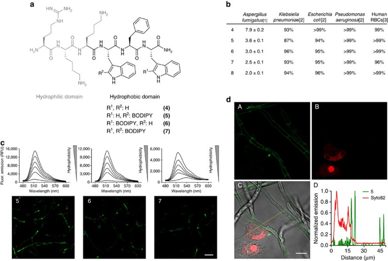
Fluorescent antimicrobial peptides are promising structures for in situ, real-time imaging of fungal infection. Here we report a fluorogenic probe to image Aspergillus fumigatus directly in human pulmonary tissue. We have developed a fluorogenic Trp-BODIPY amino acid with a spacer-free C-C linkage between Trp and a BODIPY fluorogen, which shows remarkable fluorescence enhancement in hydrophobic microenvironments. The incorporation of our fluorogenic amino acid in short antimicrobial peptides does not impair their selectivity for fungal cells, and enables rapid and direct fungal imaging without any washing steps. We have optimized the stability of our probes in human samples to perform multi-photon imaging of A. fumigatus in ex vivo human tissue. The incorporation of our unique BODIPY fluorogen in biologically relevant peptides will accelerate the development of novel imaging probes with high sensitivity and specificity.


Conflict of interest statement

The authors declare competing financial interests: University of Edinburgh has filed an invention disclosure form to protect part of the technology described in the study.

Figures

Figure 1
Figure 1. A Trp-BODIPY fluorogenic amino acid.
(a) Synthetic scheme and spectral properties of the Trp-BODIPY fluorogenic amino acid 3 (NR: no reaction). (b) The amino acid 3 displays strong fluorogenic behaviour in phospholipid membranes. Spectra of compound 3 (10 μM) were recorded after incubation with PC:cholesterol (7:1) liposome suspensions in PBS ranging from 3.75 to 0.004 mg ml−1 of PC in two-fold serial dilutions, λexc.: 450 nm. PBS alone was used as a negative control for a non-hydrophobic environment. On the right-hand side, pictures of the fluorescence emission of 3 under excitation with a 365 nm UV-lamp in PC:cholesterol liposome suspensions with increasing PC content (from top to bottom: 3.75, 1.88, 0.94, 0.47, 0.23 and 0 (only PBS) mg ml−1 of PC).
Figure 2
Figure 2. Fluorogenic peptides for live cell imaging of A.fumigatus in co-culture with human lung epithelial cells.
(a) Chemical structures of non-labelled and fluorogenic linear peptides (4-7), highlighting the two conserved hydrophilic (grey) and hydrophobic (black) domains of Peptide Antifungal 26 (PAF26). (b) Activity of antimicrobial peptides in A. fumigatus, several bacterial strains and in human RBCs.[1] IC50 (μM) values represented as means±s.e.m. from n=3, [2] cell viability upon 16 h incubation with 48 at their respective IC50 concentrations (n=3), [3] cell viability upon 1 h incubation with 48 at their respective IC50 concentrations (n=3). (c) Fluorogenic behaviour of 57 (10 μM) in phosphatidylcoline (PC):cholesterol (7:1) liposome suspensions in PBS ranging from 3.75 to 0.004 mg ml−1 of PC in two-fold serial dilutions (λexc.: 450 nm), and wash-free live cell images of A. fumigatus at 37 °C using fluorescence confocal microscopy after incubation with peptides 57 (5 μM). Scale bar, 20 μm. (d) Peptide 5 (5 μM, green) and Syto82 (2.5 μM, red counterstain for lung epithelial cells) were incubated in co-cultures of A. fumigatus and human lung A549 epithelial cells and imaged under a fluorescence confocal microscope at 37 °C without any washing steps. Fluorescence staining of 5 (A), Syto82 (B), merged (C) and plot profile analysis (D) of peptide 5 (green) and Syto82 (red) from image C. Scale bar, 10 μm.
Figure 3
Figure 3. The cyclic peptide 8 is a highly stable fluorogenic agent for high-resolution imaging of A. fumigatus.
(a) Comparative chemical stability of mono-labelled BODIPY linear (5) and cyclic (8) PAF26 analogues in human bronchoalveolar lavage samples from patients with acute respiratory distress syndrome. (b) Chemical structure of the cyclic BODIPY-labelled peptide 8. (c) Kinetic analysis (from time-lapse imaging in d of the fluorescence signal of compound 8 (2 μM) in the cell membrane of A. fumigatus (arrow points at the addition time for compound 8). (d) Time-lapse high-resolution imaging of A. fumigatus upon incubation with a cell membrane counterstain (red) and compound 8 (2 μM, green) for 0 min (i), 1 min (ii), 3 min (iii) and 10 min (iv) (see Supplementary Movie 3). Scale bar, 2.5 μm.
Figure 4
Figure 4. Multi-photon fluorescence microscopy of ex vivo human pulmonary tissue after incubation with RFP-expressing A. fumigatus.
(a) Multi-photon microscope images from peptide 8 (5 μM) (A), RFP-expressing A. fumigatus (B), second harmonic generation from collagen fibres (C) and merged (D) in ex vivo human lung tissue. Scale bar, 10 μm. (b) (A) Fluorescence lifetime image of 8-stained A. fumigatus in ex vivo human lung tissue. White arrows point autofluorescent tissue structures and yellow arrows point 8-stained fungal cells. (B) Corresponding fluorescence image of 8-stained A. fumigatus (green) and collagen fibres (second harmonic generation, cyan) for the fluorescence lifetime image in A. Scale bar, 20 μm.

References

    1. Chai L. Y. & Hsu L. Y. Recent advances in invasive pulmonary aspergillosis. Curr. Opin. Pulm. Med. 17, 160–166 (2011). - PubMed
    1. Azoulay E. & Afessa B. Diagnostic criteria for invasive pulmonary aspergillosis in critically ill patients. Am. J. Respir. Crit.Care Med. 186, 8–10 (2012). - PubMed
    1. Zhao M. et al. Spatial-temporal imaging of bacterial infection and antibiotic response in intact animals. Proc. Natl Acad. Sci. USA 98, 9814–9818 (2001). - PMC - PubMed
    1. Leevy W. M. et al. Optical imaging of bacterial infection in living mice using a fluorescent near-infrared molecular probe. J. Am. Chem. Soc. 128, 16476–16477 (2006). - PMC - PubMed
    1. Leevy W. M. et al. Noninvasive optical imaging of staphylococcus aureus bacterial infection in living mice using a Bis-dipicolylamine-Zinc(II) affinity group conjugated to a near-infrared fluorophore. Bioconjug. Chem. 19, 686–692 (2008). - PMC - PubMed

Publication types

MeSH terms