

Interleukin-31 expression and relation to disease severity in human asthma
- PMID: 26956917
- PMCID: PMC4783779
- DOI: 10.1038/srep22835
Interleukin-31 expression and relation to disease severity in human asthma
Abstract
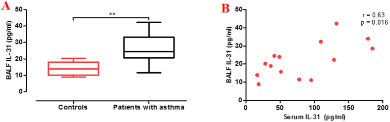
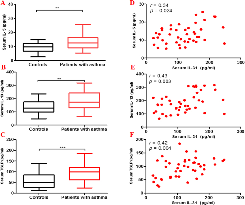
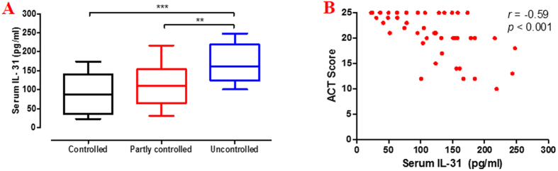
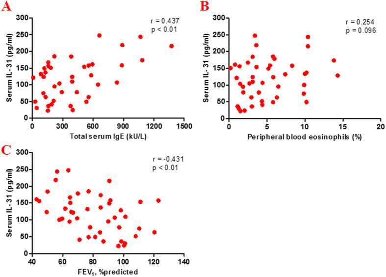
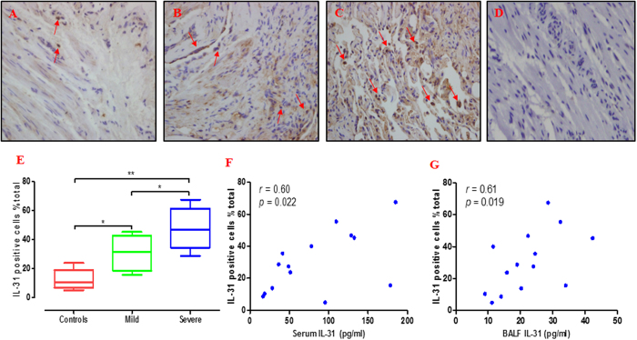
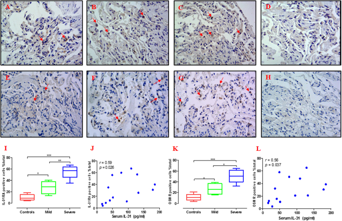
Interleukin 31 (IL-31) is a novel T helper type 2 effector cytokine that plays an important role in the pathogenesis of allergic diseases. However, its role in human asthma remains unclear. The aim of this study was to measure IL-31 levels in the serum, bronchoalveolar lavage fluid (BALF) and bronchial tissue of asthmatics and healthy subjects, and identify its possible correlation to disease severity. We quantified IL-31 levels in the serum of patients with asthma (n = 44), as well as in controls (n = 22). Of these subjects, 9 asthmatics and five controls underwent bronchoscopy with endobronchial biopsy and BALF collection. Our data showed that serum and BALF IL-31 levels were significantly elevated in patients with asthma compared with controls. Expressions of IL-31 and IL-31 receptor (IL-31RA and OSMR) were more prominent in the bronchial tissue in severe compared to mild asthma and controls. Serum IL-31 levels correlated positively with Th2 related cytokines (IL-5, IL-13, and TSLP), asthma severity or total serum immunoglobulin E (IgE), and inversely with asthma control and the forced expiratory volume in 1 second (FEV1). The current data may provide insight into the underlying pathogenesis of asthma, in which IL-31 has an important pathogenic role.
Figures

Similar articles
-
Osteopontin expression and relation to disease severity in human asthma.Eur Respir J. 2011 Feb;37(2):331-41. doi: 10.1183/09031936.00017810. Epub 2010 Jun 18. Eur Respir J. 2011. PMID: 20562127
-
Elevated Expression of IL-33 and TSLP in the Airways of Human Asthmatics In Vivo: A Potential Biomarker of Severe Refractory Disease.J Immunol. 2018 Apr 1;200(7):2253-2262. doi: 10.4049/jimmunol.1701455. Epub 2018 Feb 16. J Immunol. 2018. PMID: 29453280
-
Allergic and nonallergic asthmatics have distinct patterns of T-cell activation and cytokine production in peripheral blood and bronchoalveolar lavage.Am Rev Respir Dis. 1992 Jul;146(1):109-15. doi: 10.1164/ajrccm/146.1.109. Am Rev Respir Dis. 1992. PMID: 1626792
-
Inflammatory determinants of asthma severity: mediator and cellular changes in bronchoalveolar lavage fluid of patients with severe asthma.J Allergy Clin Immunol. 1996 Nov;98(5 Pt 2):S27-33; discussion S33-40. J Allergy Clin Immunol. 1996. PMID: 8939174 Review.
-
How to measure airway inflammation: bronchoalveolar lavage and airway biopsies.Can Respir J. 1998 Jul-Aug;5 Suppl A:18A-21A. Can Respir J. 1998. PMID: 9753511 Review.
Cited by
-
Biomarkers of type 2 and non-type 2 inflammation in asthma exacerbations.Cent Eur J Immunol. 2024;49(2):203-213. doi: 10.5114/ceji.2024.141345. Epub 2024 Jul 12. Cent Eur J Immunol. 2024. PMID: 39381551 Free PMC article.
-
Difference of Serum Cytokine Profile in Allergic Asthma Patients According to Disease Severity.J Asthma Allergy. 2022 Mar 6;15:315-326. doi: 10.2147/JAA.S345759. eCollection 2022. J Asthma Allergy. 2022. PMID: 35283636 Free PMC article.
-
IL-31 plays dual roles in lung inflammation in an OVA-induced murine asthma model.Biol Open. 2019 Jan 15;8(1):bio036244. doi: 10.1242/bio.036244. Biol Open. 2019. PMID: 30647024 Free PMC article.
-
Increased IL-31 expression in serum and tissue protein in prurigo nodularis.Ther Adv Chronic Dis. 2022 Jul 19;13:20406223221112561. doi: 10.1177/20406223221112561. eCollection 2022. Ther Adv Chronic Dis. 2022. PMID: 35875833 Free PMC article.
-
Increase In Il-31 Serum Levels Is Associated With Reduced Structural Damage In Early Axial Spondyloarthritis.Sci Rep. 2018 May 16;8(1):7731. doi: 10.1038/s41598-018-25722-z. Sci Rep. 2018. PMID: 29769586 Free PMC article.
References
-
- Cornelissen C., Lüscher-Firzlaff J., Baron J. M. & Lüscher B. Signaling by IL-31 and functional consequences. Eur J Cell Biol 91, 552–566 (2012). - PubMed
-
- Global Initiative for Asthma. GINA Report: Global strategy for asthma management and prevention (2015 Update). Available at: http://www.ginasthma.com. Accessed May, 20, 2015.
-
- Dillon S. R. et al. Interleukin 31, a cytokine produced by activated T cells, induces dermatitis in mice. Nat Immunol 5, 752–760 (2004). - PubMed
Publication types
MeSH terms
Substances
LinkOut - more resources
Full Text Sources
Other Literature Sources
Medical
