. 2016 Mar 9:7:10955.
doi: 10.1038/ncomms10955.

MICU1 regulation of mitochondrial Ca(2+) uptake dictates survival and tissue regeneration

Affiliations

MICU1 regulation of mitochondrial Ca(2+) uptake dictates survival and tissue regeneration

Anil Noronha Antony et al. Nat Commun. .

Abstract

Mitochondrial Ca(2+) uptake through the recently discovered Mitochondrial Calcium Uniporter (MCU) is controlled by its gatekeeper Mitochondrial Calcium Uptake 1 (MICU1). However, the physiological and pathological role of MICU1 remains unclear. Here we show that MICU1 is vital for adaptation to postnatal life and for tissue repair after injury. MICU1 knockout is perinatally lethal in mice without causing gross anatomical defects. We used liver regeneration after partial hepatectomy as a physiological stress response model. Upon MICU1 loss, early priming is unaffected, but the pro-inflammatory phase does not resolve and liver regeneration fails, with impaired cell cycle entry and extensive necrosis. Ca(2+) overload-induced mitochondrial permeability transition pore (PTP) opening is accelerated in MICU1-deficient hepatocytes. PTP inhibition prevents necrosis and rescues regeneration. Thus, our study identifies an unanticipated dependence of liver regeneration on MICU1 and highlights the importance of regulating MCU under stress conditions when the risk of Ca(2+) overload is elevated.


Figures

Figure 1
Figure 1. Phenotype of MICU1−/− mouse and mitochondrial Ca2+ uptake in MICU1−/− MEFs.
(a) Map of the MICU1 gene with exon 3 flanked by two loxP sites and a Neomycin selection cassette flanked by flippase recognition target sites (FRTs) and subsequent recombinations for removal of the Neomycin cassette and the exon 3, leading to the truncation of the MICU1 gene. (b) Representative images of haematoxylin and eosin (H&E) staining of diaphragm, heart and lung from MICU1+/+ and MICU1−/− animals at e18.5 show no morphological abnormalities (× 40). (c) Quantification of the neuron count in the nucleus ambiguus and the nucleus facialis in toluidine blue-stained sections of the hindbrain of MICU1+/+ and MICU1−/− e18.5 animals. Individual points are averages from different animals. Horizontal lines show mean values. Counting of neurons in the nucleus tractus solitarius that contains a subset of inspiratory neurons was not feasible because its borders were less defined. (d) Immunoblots of MICU1, MICU2, MCU and Hsp70 in MICU1+/+ and MICU1−/− MEFs lysates. Relative protein level is displayed in the bar graph; each protein was normalized to Hsp70, and expressed relative to MICU1+/+ MEFs (mean±s.e.m., n=4, *P<0.05, Student's t-test). (e) Representative [Ca2+]c time courses of the mitochondrial clearance of a 3 μM or 20 μM CaCl2 bolus (3Ca or 20Ca) in permeabilized MICU1+/+ and MICU1−/− MEFs in the presence of thapsigargin (2 μM) and CGP-37157 (20 μM). (f) [Ca2+]c dose response of the initial mitochondrial uptake (30 s after CaCl2 addition) of different Ca2+ boluses recorded as in e. The CaCl2 doses added were (in μM) 3, 7, 10, 15 and 20 (n=4 per group). A sigmoidal fit is displayed for each. (g) Double logarithmic plot of the initial rates of Ca2+ uptake against the peak [Ca2+]c. Slope of each linear fit is indicated (mean±s.e.m., n=4/group, Student's t-test).
Figure 2
Figure 2. Liver regeneration after partial hepatectomy (PHx) in MICU1 KD and Ctrl liver.
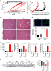
(a) Representative images of haematoxylin and eosin (H&E)-stained liver tissue and quantification of necrotic area in Ctrl and KD mice before (LL, left lateral lobe) and 30 h after PHx (Phx 30 h; collected from the same mouse for individual comparison). Magnified image displays a typical necrotic area in KD liver 30 h after PHx along with quantification. Scale bars, 200 μm (mean±s.e.m., n=4 per group). (b) Serum levels of alanine aminotransferase (ALT), direct and total bilirubin and triglycerides in Ctrl and KD mice 30 h post PHx, or after sham surgery (mean±s.e.m., n=3–6 per group). (c) BrdU immunostaining and quantification 30 h post PHx in Ctrl and KD mouse liver. Scale bars, 50 μm (mean±s.e.m., n=4 per group). (d) Immunoblotting of Cyclin D1 expression and quantification in Ctrl and KD liver lysates before PHx (LL) and 30 h post Sham operated or PHx (mean±s.e.m., n=4 per group). Values are mean±s.e.m., **P<0.005, ***P<0.0005 via Mann–Whitney test (a) and two-way analysis of variance followed by Tukey's multiple comparison test for the rest.
Figure 3
Figure 3. Early response to PHx in MICU1 KD and Ctrl liver.
(a) Immunoblot analysis of phosphorylation level of CREB and STAT3 before (LL) and 1 or 6 h after PHx. Quantification of phospho-STAT3 and phospho-CREB is displayed in bar graphs (mean±s.e.m., n=3/group). (b) mRNA fold change of genes, relative to LL, 6 h after PHx. Horizontal lines show mean values. (mean±s.e.m., n=3/group). (c) Serum level of interleukin-6 (IL-6) and tissue level of TNF-α and DNA-binding activity of NF-κB in Ctrl and KD mice 1 and 6 h after PHx (mean±s.e.m., n=3–4/group). (d) Serum levels of ALT, direct and total bilirubin, and triglycerides in Ctrl and KD mice 1 and 6 h post PHx (mean±s.e.m., n=3/group). Values are mean±s.e.m., *P<0.05, **P<0.005, ***P<0.0005 via Mann–Whitney test (c-last panel) and two-way analysis of variance followed by Tukey's multiple comparison test for the rest.
Figure 4
Figure 4. Prevention of mitochondrial Ca2+ overload rescues liver regeneration in MICU1 KD mice.
(a) Assessment of Ca2+ overload-induced mitochondrial PTP opening in Ctrl and KD permeabilized hepatocytes. Extramitochondrial [Ca2+] was recorded during repetitive addition of CaCl2 boluses (1 μM each) in the absence (Mock) or presence of RuRed (3 μM) or NIM811 (2 μM). Right panel: Ca2+ load dependence of PTP opening shown as Ca2+ efflux via PTP (Mock minus NIM811) plotted against the mitochondrial Ca2+ uptake (RuRed minus Mock), mean traces of triplicates. Representative of n=4 independent experiments. (b) Representative images of haematoxylin and eosin (H&E)-stained liver tissue 30 h post PHx showing the absence of necrotic area in both Ctrl and KD liver treated without and with 10 mg kg−1 NIM811 (4 separate mice). Liver necrosis was evident in vehicle (Veh)-treated KD mice. Scale bars, 200 μm. (c) BrdU immunostaining and quantification 30 h post PHx in Ctrl and KD mouse liver. Scale bars, 50 μm (mean±s.e.m., n=5 per group). (d) Serum levels of ALT, direct and total bilirubin and triglycerides in Ctrl and KD mice after 30 h of PHx and treated with either vehicle or NIM811. (mean±s.e.m., n=5 per group). (e) Cyclin D1 expression assessed by immunoblotting in Ctrl and KD liver lysates of Ctrl and KD mice treated with NIM811 or vehicle before (LL) and after 30 h PHx (mean±s.e.m., n=3–4/group). Values are mean±s.e.m., *P<0.05, **P<0.005, ***P<0.0005 via two-way analysis of variance followed by Tukey's multiple comparison test.
Figure 5
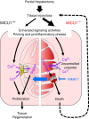
Figure 5. Loss of control over mitochondrial Ca2+ uptake turns regeneration signals to death signals in MICU1-deficient hepatocytes.
Partial hepatectomy, a surgical model for extensive tissue injury/loss in the liver, reprogrammes the fully differentiated hepatocytes in the remnant liver to proliferate in order to regenerate liver parenchyma. Reprogramming involves the synchronized priming of hepatocytes by growth factors and pro-inflammatory cytokines to obtain replicative competence. These mediators engage a broad range of intracellular signals, including Ca2+, which in addition to driving cytosolic effectors, can propagate to the mitochondrial matrix and control metabolic enzyme activities to support the proliferative response. Mitochondrial Ca2+ uptake is mediated by the Ca2+-gated mitochondrial Ca2+ uniporter complex. Ca2+ gating occurs through MICU1 (yellow trapezoid), which associates with the MCU pore (grey cylinder) from the intermembrane space side to set the threshold for Ca2+ activation. As a result, mitochondrial Ca2+ uptake occurs largely through local, high [Ca2+] elevations, whereas smaller events and global Ca2+ fluctuations are repelled (left-side purple arrows). Upon loss of MICU1 control over the Ca2+ uptake threshold, smaller [Ca2+] elevations can activate uniporter-mediated uptake (right-side purple arrows). Ca2+ signalling events occurring after PHx can thereby lead to mitochondrial Ca2+ overload to enhance permeability transition pore (PTP) opening and switch the cell's fate from proliferation to a death track. Large-scale hepatocyte death will overwhelm the functional capacity of the remaining liver cells resulting in organ failure (dashed arrow). Pharmacological inhibition of the PTP by NIM811 rescues mitochondrial competence to support proliferation even under the higher Ca2+ load in MICU1-ablated cells (thick blue arrow), which may provide the stimulus for accelerated cell proliferation under these conditions.

Comment in

References

    1. Rizzuto R., De Stefani D., Raffaello A. & Mammucari C. Mitochondria as sensors and regulators of calcium signalling. Nat. Rev. Mol. Cell. Biol. 13, 566–578 (2012). - PubMed
    1. Szabadkai G. & Duchen M. R. Mitochondria: the hub of cellular Ca2+ signaling. Physiology (Bethesda) 23, 84–94 (2008). - PubMed
    1. Kirichok Y., Krapivinsky G. & Clapham D. E. The mitochondrial calcium uniporter is a highly selective ion channel. Nature 427, 360–364 (2004). - PubMed
    1. Gunter T. E. & Pfeiffer D. R. Mechanisms by which mitochondria transport calcium. Am. J. Physiol. 258, C755–C786 (1990). - PubMed
    1. Baughman J. M. et al. Integrative genomics identifies MCU as an essential component of the mitochondrial calcium uniporter. Nature 476, 341–345 (2011). - PMC - PubMed

Publication types

Substances