. 2016 Mar 9:6:22490.
doi: 10.1038/srep22490.

In vivo reprogramming reactive glia into iPSCs to produce new neurons in the cortex following traumatic brain injury

Affiliations

In vivo reprogramming reactive glia into iPSCs to produce new neurons in the cortex following traumatic brain injury

Xiang Gao et al. Sci Rep. .

Abstract

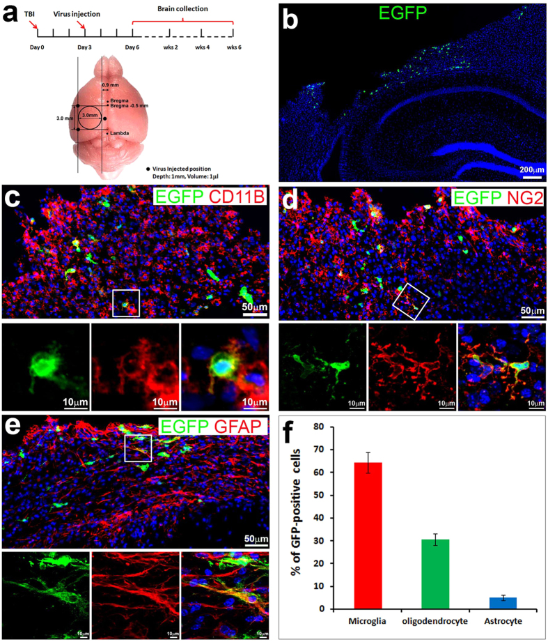
Traumatic brain injury (TBI) results in a significant amount of cell death in the brain. Unfortunately, the adult mammalian brain possesses little regenerative potential following injury and little can be done to reverse the initial brain damage caused by trauma. Reprogramming adult cells to generate induced pluripotent stem cell (iPSCs) has opened new therapeutic opportunities to generate neurons in a non-neurogenic regions in the cortex. In this study we showed that retroviral mediated expression of four transcription factors, Oct4, Sox2, Klf4, and c-Myc, cooperatively reprogrammed reactive glial cells into iPSCs in the adult neocortex following TBI. These iPSCs further differentiated into a large number of neural stem cells, which further differentiated into neurons and glia in situ, and filled up the tissue cavity induced by TBI. The induced neurons showed a typical neuronal morphology with axon and dendrites, and exhibited action potential. Our results report an innovative technology to transform reactive glia into a large number of functional neurons in their natural environment of neocortex without embryo involvement and without the need to grow cells outside the body and then graft them back to the brain. Thus this technology offers hope for personalized regenerative cell therapies for repairing damaged brain.


Figures

Figure 1
Figure 1. Retrovirus-infected reactive glia in cortex following moderate traumatic brain injury (TBI).
(a) Strategy to induce pluripotent stem cells (iPSCs) in vivo. (b) 3 days after retrovirus injection, cells around the injured area were infected and started to express enhanced green fluorescent protein (EGFP). (c) EGFP-positive cells (green) colabeled with microglia specific marker, cd11b (red). (d) EGFP-positive cells (green) colabeled with oligodendrocyte progenitor cell marker, neural/glial antigen 2 (NG2, red). (e) EGFP-positive cells co-expressed astrocyte marker, glial fibrillary acidic protein (GFAP, red). (f) Quantification to show the ratio of EGFP-positive cells co-expressing different glia markers.
Figure 2
Figure 2. In vivo reprogramming reactive glia to embryonic stem-like cells.
Tracing the fates of retrovirus infected cells in the injured cortex 2 weeks and 4 weeks after traumatic brain injury (TBI) with immunostaining. (a) Cells expressing EGFP (green) around the injury area 2 weeks after TBI. (b) Cell clusters (white arrows) expressing 4 human transcriptional factors (hOCT4, hKLF4, hSOX2 and hcMYC, hOKSM) and EGFP (green) around the injury area 2 weeks after TBI. (c) Cells expressing hOKSM-EGFP (green) also colabeled with Nanog (red). c1–4. Images of single focal section images to show colabeling of hOKSM/EGFP (green) with Nanog (red) in the cells within the white box from panel (c). (d–g) hOKSM/EGFP (green) expressing cells form cluster and expressed SSEA4 4 weeks after TBI. (h(x–z)). Images of three-dimensional reconstruction to confirm that a hOKSM/EGFP expressing cell in the white box of panel (g) also expressed SSEA4.
Figure 3
Figure 3. Reprogrammed glia produced cells in all three embryonic germ layers at 4 weeks after TBI.
Cells expressing hOKSM-EGFP expanded into cellular clusters and expressed markers of all three embryonic germ layers in the injured cortex at 4 weeks after traumatic brain injury (TBI). (a–d) A large number of hOKSM-EGFP expressing cells (green) in the clusters highly expressed Sox2 (red), a marker for embryonic ectoderm. (e–h) Images of single focal section to show colabeling of hOKSM-EGFP (green) with Sox2 (red) in the cells within the white box from panel (d). (i) A small number of the hOKSM-EGFP expressing cells expanded into cell clusters and expressed brachyury, a marker of embryonic mesoderm. White circles indicate neural tube-like structures formed by EGFP-positive cells. (j–m) Enlarged images of cells within the white box in the panel (i). (n(x–z)) Images of three-dimensional reconstruction to confirm that some of the hOKSM-EGFP expressing cells in the white box of panel (i) also expressed Brachyary. (o) A small number of the hOKSM-EGFP expressing cells expanded into cell clusters and expressed Gata4, a marker of embryonic endoderm. (p–s) Enlarged images of cells within the white box in the panel (o). White circles indicate neural tube-like structures formed by EGFP-positive cells. Yellow arrowheads pointed out the reprogrammed cells with long processes.
Figure 4
Figure 4. Reprogrammed cells differentiated into neural fate at 4 weeks after traumatic brain injury (TBI).
(a) hOKSM-EGFP expressing cells formed neural tube-like structures (white circle) in the injured cortex. (b–e) EGFP-positive cell cluster (EGFP) exhibited typical neural tube-like structure and expressed Nestin (red), a marker used for neural stem cells. (f–h) Images of three-dimensional reconstruction to confirm that a hOKSM-EGFP expressing cell in the panel e also expressed Nestin. (j) Some of the hOKSM-EGFP expressing cells (green) started to differentiate into doublecortin (Dcx)-positive immature neurons (red). (k–n) Enlarged images of cells in the white box of panel (j) to show the hOKSM-EGFP expressing cells also expressed Dcx, a marker for immature neurons.
Figure 5
Figure 5. Reprogrammed cell-derived immature neurons developed into mature neurons at 6 weeks after traumatic brain injury (TBI).
(a) The hOKSM-EGFP expressing cells (green) differentiated into a large number of mature neurons expressing neuronal nuclei protein (NeuN, red), a marker for mature neurons, in the injured cortex. (b–e) Enlarged images from pane a to show hOKSM-EGFP expressing cells (green) colabeled with NeuN (red). (f) Enlarged image from pane (e) to show individual neurons with long processes expressing both EGFP (green) and NeuN (red). (g–j) Images of three-dimensional reconstruction to confirm that a hOKSM-EGFP expressing cell in the panel (f) also expressed NeuN.
Figure 6
Figure 6. Functional integration of iNeurons.
(a–d) Confocal images of recorded EGFP-positive cells, which were also loaded with biocytin during recording. (e–g) Electrophysiology of a EGFP-positive cell labeled in panel (a). This cell fired repetitive action potentials in response to depolarizing currents (e) and showed spontaneous synaptic currents when voltage clamped at the resting membrane potential (f,g).
Figure 7
Figure 7. Reprogrammed cells also differentiate into glia at 6 weeks after traumatic brain injury (TBI).
(a–d) Some of the hOKSM-EGFP expressing cells (green) differentiated into astrocytes expressing GFAP (red) in the injured cortex at 6 weeks after TBI. (e–h) Images of three-dimensional reconstruction to confirm that a hOKSM-EGFP expressing cell in the panel (d) also expressed GFAP. (i) A subpopulation of the hOKSM-EGFP expressing cells (green) differentiated into oligodendrocyte precursors expressing NG2 (red) in the injured cortex. (j) Images of three-dimensional reconstruction to confirm that a hOKSM-EGFP expressing cell in the panel (i) also expressed NG2.

References

    1. Langlois J. A., Rutland-Brown W. & Thomas K. E. The incidence of traumatic brain injury among children in the United States: differences by race. J Head Trauma Rehabil 20, 229–238 (2005). - PubMed
    1. Minino A. M., Anderson R. N., Fingerhut L. A., Boudreault M. A. & Warner M. Deaths: injuries, 2002. Natl Vital Stat Rep 54, 1–124 (2006). - PubMed
    1. Hoge C. W., Goldberg H. M. & Castro C. A. Care of war veterans with mild traumatic brain injury–flawed perspectives. N Engl J Med 360, 1588–1591, 360/16/1588 (2009). - PubMed
    1. Hoge C. W. et al. Mild traumatic brain injury in US Soldiers returning from Iraq. N Engl J Med 358, 453–463, NEJMoa072972 (2008). - PubMed
    1. Stiles J., Reilly J., Paul B. & Moses P. Cognitive development following early brain injury: evidence for neural adaptation. Trends Cogn Sci 9, 136–143 (2005). - PubMed

Publication types