. 2016 May 6;291(19):10046-57.
doi: 10.1074/jbc.M115.711762. Epub 2016 Mar 8.

Structural Basis for Translocation of a Biofilm-supporting Exopolysaccharide across the Bacterial Outer Membrane

Affiliations

Structural Basis for Translocation of a Biofilm-supporting Exopolysaccharide across the Bacterial Outer Membrane

Yan Wang et al. J Biol Chem. .

Abstract

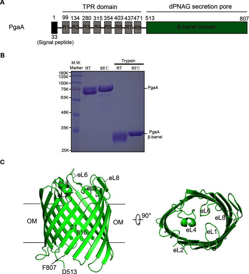
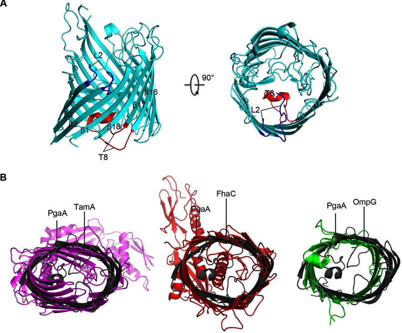
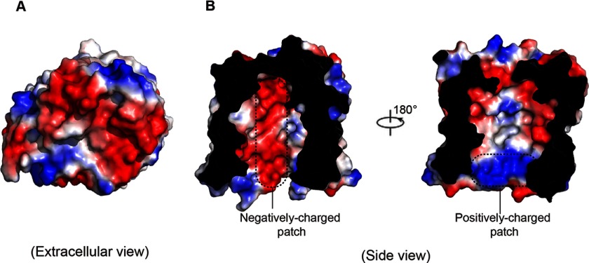
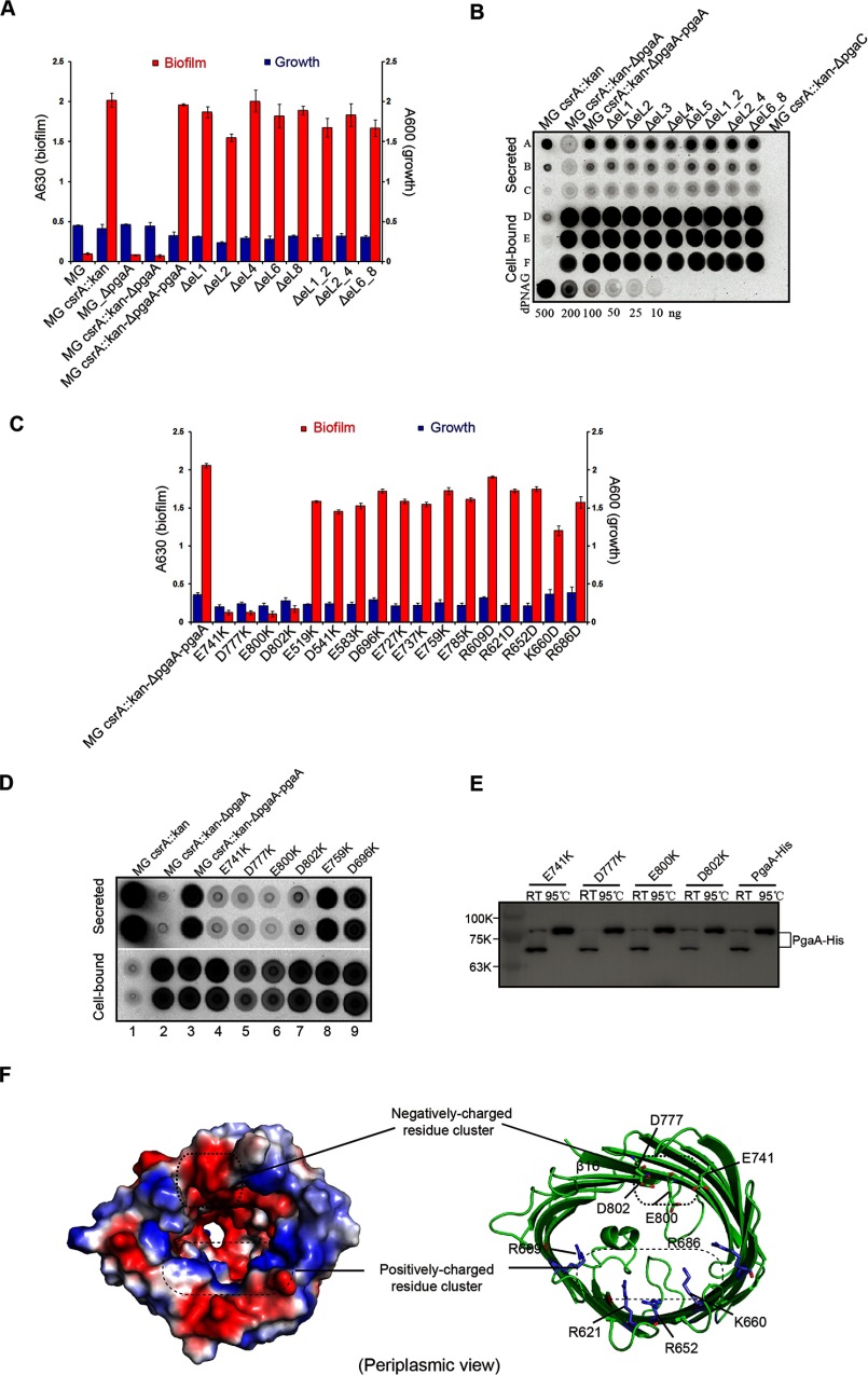
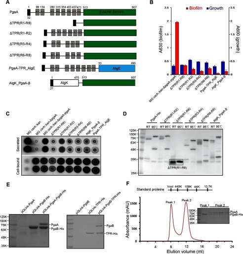
The partially de-N-acetylated poly-β-1,6-N-acetyl-d-glucosamine (dPNAG) polymer serves as an intercellular biofilm adhesin that plays an essential role for the development and maintenance of integrity of biofilms of diverse bacterial species. Translocation of dPNAG across the bacterial outer membrane is mediated by a tetratricopeptide repeat-containing outer membrane protein, PgaA. To understand the molecular basis of dPNAG translocation, we determined the crystal structure of the C-terminal transmembrane domain of PgaA (residues 513-807). The structure reveals that PgaA forms a 16-strand transmembrane β-barrel, closed by four loops on the extracellular surface. Half of the interior surface of the barrel that lies parallel to the translocation pathway is electronegative, suggesting that the corresponding negatively charged residues may assist the secretion of the positively charged dPNAG polymer. In vivo complementation assays in a pgaA deletion bacterial strain showed that a cluster of negatively charged residues proximal to the periplasm is necessary for biofilm formation. Biochemical analyses further revealed that the tetratricopeptide repeat domain of PgaA binds directly to the N-deacetylase PgaB and is critical for biofilm formation. Our studies support a model in which the positively charged PgaB-bound dPNAG polymer is delivered to PgaA through the PgaA-PgaB interaction and is further targeted to the β-barrel lumen of PgaA potentially via a charge complementarity mechanism, thus priming the translocation of dPNAG across the bacterial outer membrane.

Keywords: PgaA; bacterial adhesion; biofilm; exopolysaccharide; extracellular matrix; membrane protein; outer membrane; outer membrane protein; poly-β-1,6-N-acetyl-d-glucosamine; polysaccharide; translocation.


Figures

FIGURE 1.
FIGURE 1.
Molecular structure of PgaA-(513–807). A, schematic structure of the full-length PgaA. Residues 1–33 constitute a signal peptide. Eight TPR motifs are denoted as R1-R8. The C terminus of PgaA (residues 513–807) forms a transmembrane β barrel. B, the full-length PgaA protein purified from membrane fraction and its trypsin-digested product. Both the full-length PgaA and PgaA β-barrel exhibit heat-modifiable mobility to certain degree on 12% SDS-PAGE. RT, room temperature. C, ribbon representation of the structure of PgaA-(513–807). The PgaA β-barrel consists of 16 β-strands, closed on the extracellular surface by four long extracellular loops eL1, eL4, eL6. and eL8. OM, outer membrane.
FIGURE 2.
FIGURE 2.
Structural comparison between PgaA-(513–807) and other representative β-barrel outer membrane proteins. A, ribbon representation of the structure of AlgE (PDB code 3RBH). AlgE β-barrel consists of 18 β-strands and the lumen is occluded by two functionally important loops: a long periplasmic loop 8 (T8, in red) and an extracellular loop 2 (L2, in blue). B, structural overlay between PgaA β-barrel and that of TamA (PDB code 4C00), FhaC (PDB code 3NJT), or OmpG (PDB code 2F1C).
FIGURE 3.
FIGURE 3.
Electrostatic potential surface representation of PgaA-(513–807). A, electrostatic potential surface representation showing that the extracellular surface of PgaA is primarily electronegative. B, a slab view showing the electrostatic properties inside of the lumen of PgaA. The surface is colored according to electrostatic potential, ranging from blue (+70 kT/e) to red (−70 kT/e), where kT is thermal energy, and e is the elementary charge. The characteristic negatively charged patch and the positively charged patch inside of the β barrel are indicated with dotted lines.
FIGURE 4.
FIGURE 4.
A cluster of negatively charged residues inside of the barrel and in proximity to periplasm is important for dPNAG secretion. A, extracellular loops of PgaA are not important for biofilm production. Wild-type E. coli strain MG1655 (denoted as MG) produced low levels of biofilm due to repression by CsrA (A630 < 0.1); the csrA-disrupted E. coli strain (denoted as MG_csrA::kan) produced 20-fold more biofilm (A630 = 2.0); both the pgaA-deleted E. coli strain (denoted as MG_ΔpgaA) and E. coli strain MG1655 with both csrA- and pgaA-disrupted (denoted as MG_csrA::kanpgaA) produced low levels of biofilm (A630 < 0.1). When MG_csrA::kanpgaA was complemented with a plasmid that expresses PgaA (MG_csrA::kanpgaA_pgaA), biofilm production was restored. None of the loop deletion mutants affected biofilm production. B, extracellular loops of PgaA are not important for secretion of dPNA. Immunoblotting of secreted and cell-bound dPNAG from MG1655 csrA::kan_ΔpgaA strain was complemented with plasmids expressing wild-type pgaA or mutant pgaA proteins with extracellular loop deletions. Secreted and cell-bound dPNAG samples were blotted onto a nitrocellulose membrane and detected with α-PNAG monoclonal antibody as described (14). Rows A–C, dPNAG derived from 30, 6, and 2 μl of spent medium. Rows D-F, cell-bound dPNAG from 40, 4, and 1 ml of culture. Dilutions of purified dPNAG (bottom row) served as a standard for comparison. C, 12 negatively charged residues (Glu-519, Asp-541, Glu-583, Asp-696, Glu-727, Glu-737, Glu-741, Asp-759, Asp-777, Glu-785, Glu-800, and D802) and 5 positively charged residues (Arg-609, Arg-621, Arg-652, Lys-660, and Arg-686) inside the lumen were mutated to Lys and Asp, respectively, and their effects on biofilm production were assayed. Each experiment was conducted at least twice in replicates of three. Error bars denote S.D. D, immunoblotting of secreted and cell-bound dPNAG from MG_csrA:: kan_ΔpgaA strains expressing wild-type PgaA or single-point PgaA mutants. Duplicate samples represent dPNAG extracted from two independent cultures. E, nickel column purification and Western blot analysis of the expression levels of both the wild-type and mutant proteins from bacterial outer membrane. Both the wild-type and mutant proteins exhibited apparent heat-modifiable mobility, suggesting that they are all folded and resided in the outer membrane. RT, room temperature. F, residues Glu-741, Asp-777, Glu-800, and Asp-802 of PgaA form a negatively charged residue cluster in proximity to the periplasm.
FIGURE 5.
FIGURE 5.
The TPR domain of PgaA directly binds PgaB and is necessary for biofilm production. A, schematic structures of the full-length PgaA, TPR-deletion mutant PgaA proteins, and two fusion proteins (AlgK_PgaA-β and PgaA-TPR_AlgE). B, TPR domain of PgaA is required for biofilm production. TPR repeat deletion mutants ΔTPR(R1-R8) (Mr 35.1), ΔTPR(R1-R2) (Mr 76.3), ΔTPR(R3-R4) (Mr 73.5), and ΔTPR(R6-R8) (Mr 73.8) all significantly affected biofilm production; two fusion proteins AlgK_PgaA-β (Mr 76.3) and PgaA-TPR_AlgE (Mr 89.3) are unable to restore biofilm production. Each experiment was conducted at least twice in replicates of three. Error bars denote S.D. C, immunoblotting of secreted and cell-bound dPNAG from MG_csrA::kan_ΔpgaA strain expressing wild-type PgaA or PgaA with TPR deletions or PgaA fusion proteins. D, nickel-column purification and Western blot analysis of the expression levels of the wild-type, various TPR-deletion PgaA mutant proteins, and fusion protein AlgK_PgaA-β-barrel from bacterial outer membranes. All the proteins exhibited apparent heat-modifiable mobility, suggesting that they are all folded and resided in the outer membranes. RT, room temperature. E, the TPR domain of PgaA directly binds PgaB. When co-expressed in bacteria, PgaB-His is capable of pulling down PgaA (left panel), and TPR-His is capable of pulling down PgaB (right panel). Pulldown mixtures were resolved on 12% SDS-PAGE gels and stained with Coomassie Blue dye. F, size exclusion chromatographic profile of the pulldown mixture of PgaB-His and PgaA on a Superdex 200 10/30 column. PgaB-His (a lipoprotein) alone is not stable and forms aggregates in a gel filtration buffer that contains 20 mm Tris, pH 8.0, 150 mm NaCl, and 0.06% n-dodecyl-β-d-maltoside, so excess PgaB-His protein was eluted in a void volume (about 8.0 ml). PgaB-His and PgaA seem to form a 1:1 stable complex as resolved by 12% SDS-PAGE analysis. mAU, milliabsorption units.

References

    1. López D., Vlamakis H., and Kolter R. (2010) Biofilms. Cold Spring Harb. Perspect. Biol. 2, a000398. - PMC - PubMed
    1. Branda S. S., Vik S., Friedman L., and Kolter R. (2005) Biofilms: the matrix revisited. Trends Microbiol. 13, 20–26 - PubMed
    1. Flemming H. C., and Wingender J. (2010) The biofilm matrix. Nat. Rev. Microbiol, 8, 623–633 - PubMed
    1. Mah T. F., and O'Toole G. A. (2001) Mechanisms of biofilm resistance to antimicrobial agents. Trends Microbiol. 9, 34–39 - PubMed
    1. Donlan R. M., and Costerton J. W. (2002) Biofilms: survival mechanisms of clinically relevant microorganisms. Clin. Microbiol. Rev. 15, 167–193 - PMC - PubMed

Publication types

MeSH terms

Substances

LinkOut - more resources