. 2016 Apr;67(9):2587-601.
doi: 10.1093/jxb/erw082. Epub 2016 Mar 8.

The unique structural and biochemical development of single cell C4 photosynthesis along longitudinal leaf gradients in Bienertia sinuspersici and Suaeda aralocaspica (Chenopodiaceae)

Affiliations

The unique structural and biochemical development of single cell C4 photosynthesis along longitudinal leaf gradients in Bienertia sinuspersici and Suaeda aralocaspica (Chenopodiaceae)

Nuria K Koteyeva et al. J Exp Bot. 2016 Apr.

Abstract

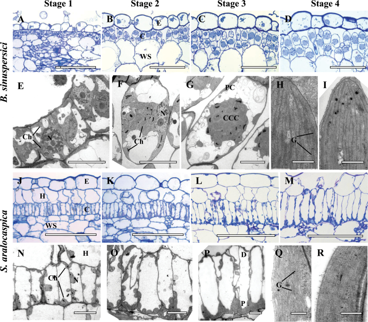
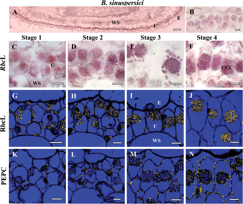
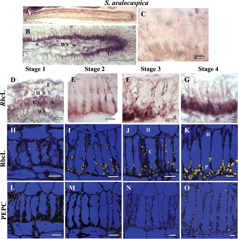
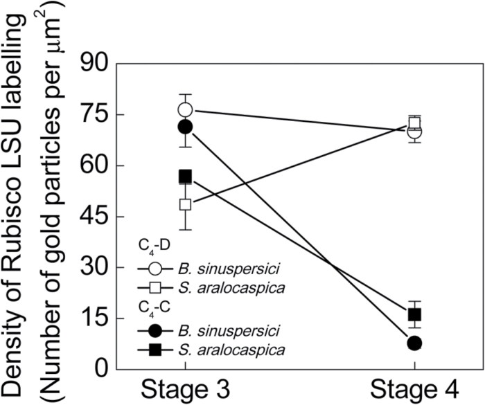
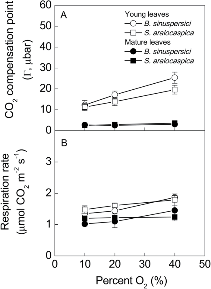
Temporal and spatial patterns of photosynthetic enzyme expression and structural maturation of chlorenchyma cells along longitudinal developmental gradients were characterized in young leaves of two single cell C4 species, Bienertia sinuspersici and Suaeda aralocaspica Both species partition photosynthetic functions between distinct intracellular domains. In the C4-C domain, C4 acids are formed in the C4 cycle during capture of atmospheric CO2 by phosphoenolpyruvate carboxylase. In the C4-D domain, CO2 released in the C4 cycle via mitochondrial NAD-malic enzyme is refixed by Rubisco. Despite striking differences in origin and intracellular positioning of domains, these species show strong convergence in C4 developmental patterns. Both progress through a gradual developmental transition towards full C4 photosynthesis, with an associated increase in levels of photosynthetic enzymes. Analysis of longitudinal sections showed undeveloped domains at the leaf base, with Rubisco rbcL mRNA and protein contained within all chloroplasts. The two domains were first distinguishable in chlorenchyma cells at the leaf mid-regions, but still contained structurally similar chloroplasts with equivalent amounts of rbcL mRNA and protein; while mitochondria had become confined to just one domain (proto-C4-D). The C4 state was fully formed towards the leaf tips, Rubisco transcripts and protein were compartmentalized specifically to structurally distinct chloroplasts in the C4-D domains indicating selective regulation of Rubisco expression may occur by control of transcription or stability of rbcL mRNA. Determination of CO2 compensation points showed young leaves were not functionally C4, consistent with cytological observations of the developmental progression from C3 default to intermediate to C4 photosynthesis.

Keywords: Bienertia sinuspersici; C4 plants; CO2 compensation point; Suaeda aralocaspica; development; immunolocalization; in situ hybridization; leaf anatomy; single cell C4 photosynthesis; ultrastructure..


Figures

Fig. 1.
Fig. 1.
Cleared young leaves of Bienertia sinuspersici (A, B) and Suaeda aralocaspica (C, D) viewed under UV light at different stages of vein initiation. There is acropetal formation of the central vein towards the leaf tip (illustrated by arrow in panel A) and basipetal direction of lateral vascular vein development from the tip to base of the leaf (illustrated by arrows in panel B). Scale bars: 250 μm for A, B; 500 μm for C, D.
Fig. 2.
Fig. 2.
Light and electron microscopy of Bienertia sinuspersici (A–I) and Suaeda aralocaspica (J–R) with longitudinal sections of young leaves. The sections show a basipetal developmental gradient with gradual structural differentiation of SC-C4 chlorenchyma cells with four stages along the longitudinal gradient: Stage 1 (A, E, J, N), Stage 2 (B, F, K, O), Stage 3 (C, G, L, P) and Stage 4 (D, H, I, M, Q, R). Panels A–D from B. sinuspersici and J–M from S. aralocaspica are light microscopy micrographs of longitudinal sections showing the development of chlorenchyma cell lineages from the base (A, J) to the tip (D, M) of young leaves, with the direction of maturation from left to right. Panels E–G from B. sinuspersici and N–P from S. aralocaspica, are TEM micrographs showing internal structural development within a single chlorenchyma cell along the longitudinal gradient from the base (E, N) to the middle region (G, P) of a young leaf. Panels H, I, show the ultrastructure of chloroplasts in the central cytoplasmic compartment, CCC (panel H) and the periphery (panel I) within a single chlorenchyma cell at the tip of a young B. sinuspersici leaf. Panels Q, R show ultrastructure of chloroplasts within the proximal (Q) and distal (R) regions of chlorenchyma cell at the tip of a young S. aralocaspica leaf. E, epidermis; C, chlorenchyma; CCC, central cytoplasmic compartment; D, distal end; G, grana; H, hypoderm; N, nucleus; P, proximal end; WS, water storage. Scale bars: 100 μm for A–D and J–M; 10 μm for E–G and N–P; 0.5μm for H, I, Q, R. (This figure is available in color at JXB online.)
Fig. 3.
Fig. 3.
In situ hybridization of rbcL mRNA (A–F) and in situ immunolocalization of Rubisco rbcL (G–J) and PEPC (K–N) with longitudinal sections of young leaves from base to tip of Bienertia sinuspersici at four stages of development: Stage 1 (C, G, K), Stage 2 (D, H, L), Stage 3 (E, I, M) and Stage 4 (F, J, N). The dark purple signal indicates the specific hybridization to an antisense mRNA probe for rbcL mRNA (panels A, C–F). Yellow particles (panels G–N) indicate labeling with rbcL and PEPC antibodies. (A) Basipetal gradient of rbcL transcript accumulation from base (left) to tip (right). (B) Sense probe control showing the very low background staining that occurred in mRNA sense strand hybridization reactions. E, epidermis; CCC, central cytoplasmic compartment; C, chlorenchyma; WS, water storage. Scale bars: 100 μm for A; 10 μm for B–N.
Fig. 4.
Fig. 4.
In situ hybridization of rbcL mRNA (A–G) and in situ immunolocalization of Rubisco rbcL (H–K) and PEPC (L–O) with longitudinal leaf sections from base to tip of Suaeda aralocaspica at four stages of development: Stage 1 (D, H, L), Stage 2 (E, I, M), Stage 3 (F, J, N) and Stage 4 (G, K, O). The dark purple signal indicates the specific hybridization to an antisense mRNA probe for rbcL mRNA (A, B, D–G). Yellow particles indicate peptide antibody labeling (H–O). (A, B) Basipetal gradient of rbcL transcript accumulation from base (left) to tip (right). (C) Sense probe control showing the very low background staining that occurs in mRNA sense strand hybridization reactions. E, epidermis; C, chlorenchyma; H, hypoderm; WS, water storage. Scale bars: 200 μm for A, 100 μm for B; 10 μm for C–O.
Fig. 5.
Fig. 5.
Quantitative graphical representation showing the density of immunolabeling for Rubisco rbcL in peripheral (●) versus CCC chloroplasts (○) in Bienertia sinuspersici, and in distal (■) versus proximal (□) chloroplasts of Suaeda aralocaspica at Stages 3 and 4 of development in young leaves. The y axis represents the number of gold particles per μm2 of chloroplast, and the x axis represents the developmental stages. 10–15 cell areas were used for counting in each cell type and for each stage of development.
Fig. 6.
Fig. 6.
Western blot analysis showing accumulation of C4 enzymes, Rubisco rbcL and rbcS in protein extracts taken along the length of young leaf, and from mature leaves in Bienertia sinuspersici and Suaeda aralocaspica. Total soluble proteins were extracted from young leaves (0.5–0.7cm long) divided into three sections (base, middle, tip), and from mature fully expanded leaves. Blots were probed with antibodies raised against PEPC, PPDK, NAD-ME and Rubisco rbcL and rbcS, respectively. Top: Representative western blots showing detection of each protein with the antibody indicated. Numbers listed at the right indicate molecular mass in kilodaltons. Bottom panels: Quantitative representation of western blot data taking relative intensity of labeling of mature leaf as 100%. YL, young leaf; ML, mature leaf.
Fig. 7.
Fig. 7.
CO2 compensation points (Г) measured in the light, and rates of dark respiration at variable oxygen levels for mature (filled symbols) and young (open symbols) leaves of Bienertia sinuspersici (circles) and Suaeda aralocaspica (squares).
Fig. 8.
Fig. 8.
Illustrations of major differences during development of C4 photosynthesis from the basal region to the tip of young leaves in SC-C4 species versus Kranz-type C4 species in subfamily Suaedoideae. Progressive changes in differentiation in two types of SC-C4 species (B. sinuspersici and S. aralocaspica) are summarized from the current study, and compared with two structural forms of Kranz-type Suaeda species (S. taxifolia and S. eltonica) described previously (Koteyeva et al., 2011). The numbers within the leaves refer to stages of chlorenchyma development along young leaves (length ~0.6cm). For comparison with SC-C4, the results with Kranz-type by Koteyeva et al. (2011) are summarized through four stages of development. The horizontal arrows point to specific defined changes that occur at each of the four stages. The vertical arrows indicate continual changes that occur along the longitudinal gradient. BS, bundle sheath; chp, chloroplasts; M, mesophyll; mito, mitochondria; SC-C4, single cell C4; C4-D domain, C4 cycle decarboxylation domain. (This figure is available in color at JXB online.)

References

    1. Akhani H, Barroca J, Koteeva N, Voznesenskaya E, Franceschi V, Edwards G, Ghaffari SM, Ziegler H. 2005. Bienertia sinuspersici (Chenopodiaceae): a new species from Southwest Asia and discovery of a third terrestrial C4 plant without Kranz anatomy. Systematic Botany 30, 290–301.
    1. Aubry S, Kelly S, Kümpers BMC, Smith-Unna RD, Hibberd JM. 2014. Deep evolutionary comparison of gene expression identifies parallel recruitment of trans-factors in two independent origins of C4 photosynthesis. PLOS Genetics 10, e1004365. - PMC - PubMed
    1. Bowman S, Patel M, Yerramsetty P, Mure C, Zielinski A, Bruenn J, Berry J. 2013. A novel RNA binding protein affects rbcL gene expression and is specific to bundle sheath chloroplasts in C4 plants. BMC Plant Biology 13, 138. - PMC - PubMed
    1. Brooks A, Farquhar GD. 1985. Effect of temperature on the CO2/O2 specificity of ribulose-1,5-bisphosphate carboxylase/oxygenase and the rate of respiration in the light. Planta 165, 397–406. - PubMed
    1. Chuong SDX, Franceschi VR, Edwards GE. 2006. The cytoskeleton maintains organelle partitioning required for single-cell C4 photosynthesis in Chenopodiaceae species. The Plant Cell 18, 2207–2223. - PMC - PubMed

Publication types

MeSH terms

Substances