. 2016 Mar 29;113(13):3639-44.
doi: 10.1073/pnas.1525637113. Epub 2016 Mar 8.

Human symbionts inject and neutralize antibacterial toxins to persist in the gut

Affiliations

Human symbionts inject and neutralize antibacterial toxins to persist in the gut

Aaron G Wexler et al. Proc Natl Acad Sci U S A. .

Abstract

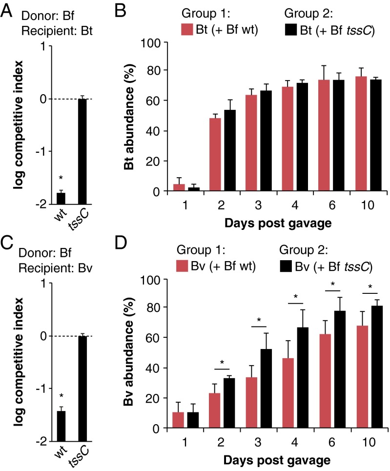
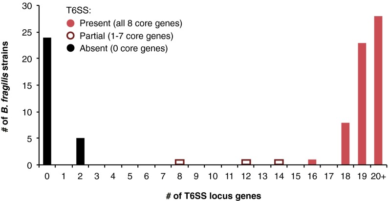
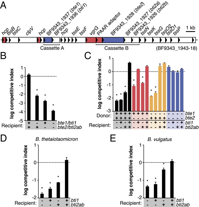
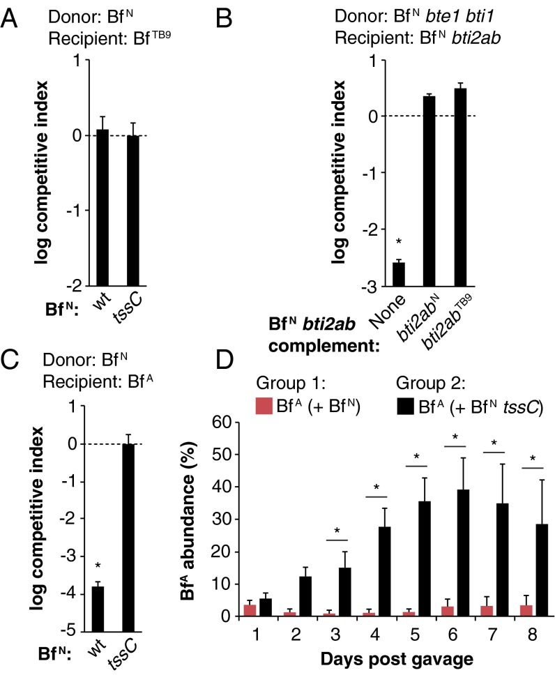
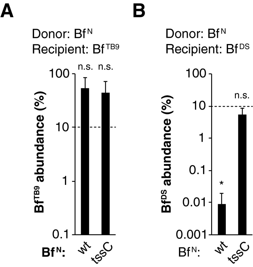
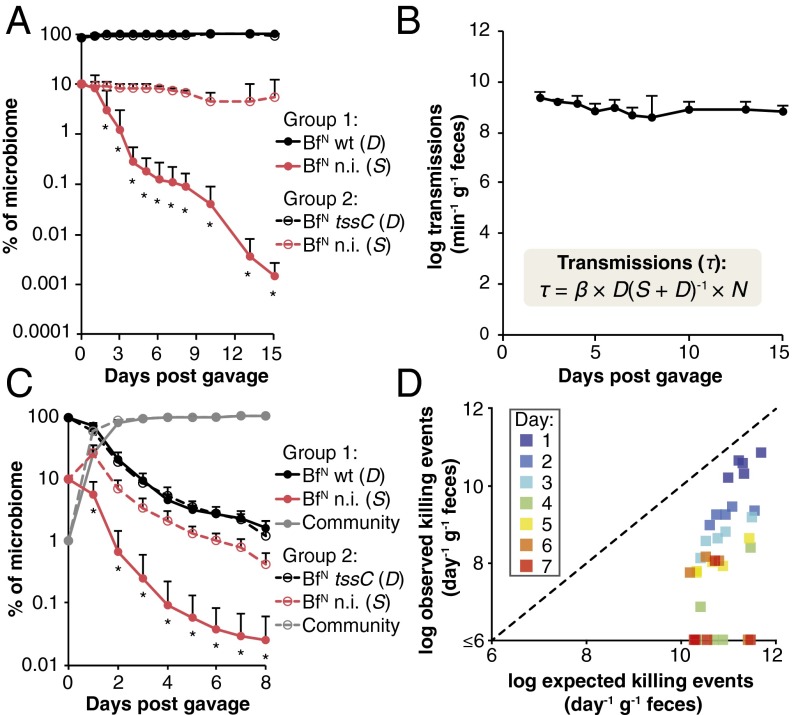
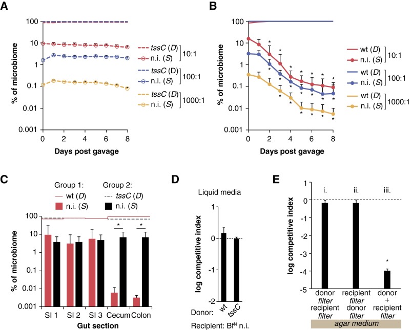
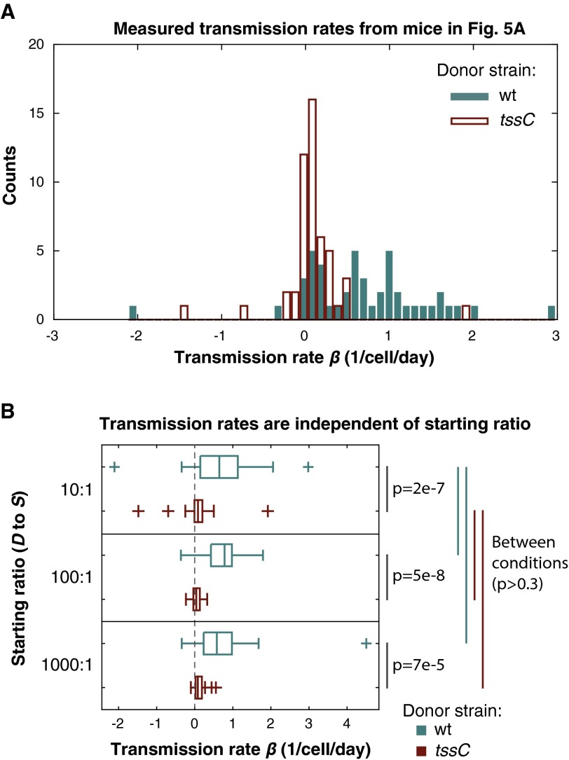
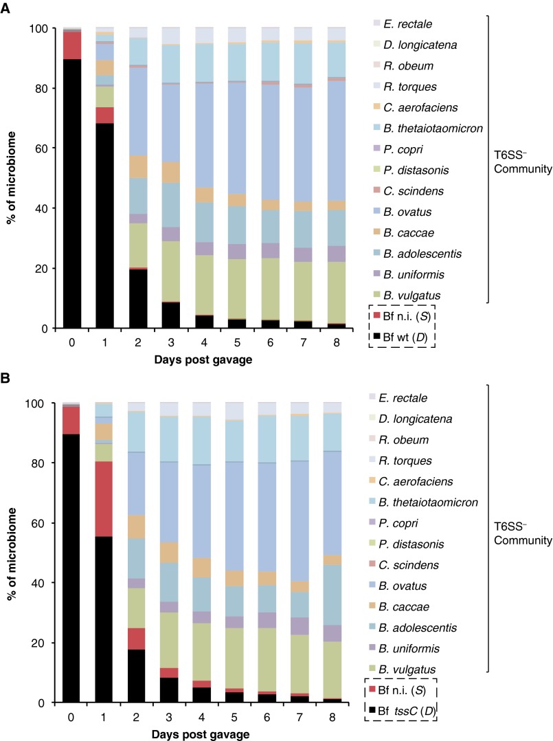
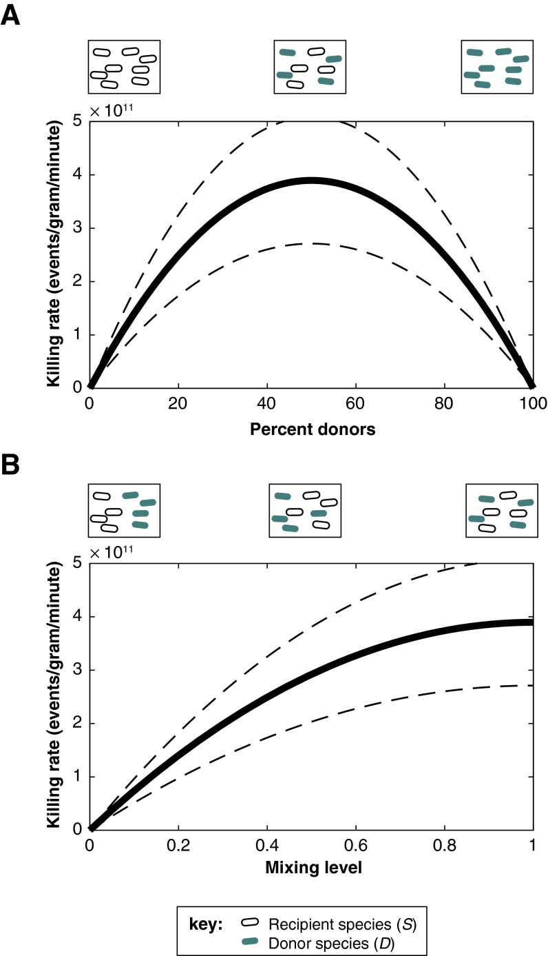
The human gut microbiome is a dynamic and densely populated microbial community that can provide important benefits to its host. Cooperation and competition for nutrients among its constituents only partially explain community composition and interpersonal variation. Notably, certain human-associated Bacteroidetes--one of two major phyla in the gut--also encode machinery for contact-dependent interbacterial antagonism, but its impact within gut microbial communities remains unknown. Here we report that prominent human gut symbionts persist in the gut through continuous attack on their immediate neighbors. Our analysis of just one of the hundreds of species in these communities reveals 12 candidate antibacterial effector loci that can exist in 32 combinations. Through the use of secretome studies, in vitro bacterial interaction assays and multiple mouse models, we uncover strain-specific effector/immunity repertoires that can predict interbacterial interactions in vitro and in vivo, and find that some of these strains avoid contact-dependent killing by accumulating immunity genes to effectors that they do not encode. Effector transmission rates in live animals can exceed 1 billion events per minute per gram of colonic contents, and multiphylum communities of human gut commensals can partially protect sensitive strains from these attacks. Together, these results suggest that gut microbes can determine their interactions through direct contact. An understanding of the strategies human gut symbionts have evolved to target other members of this community may provide new approaches for microbiome manipulation.

Keywords: gut microbiome; microbial ecology; symbiosis; type VI secretion.


Conflict of interest statement

The authors declare no conflict of interest.

Figures

Fig. 1.
Fig. 1.
T6S antagonism in the gut is species-dependent. (A and B) B. thetaiotaomicron (Bt) is susceptible to B. fragilisN (Bf) T6S in vitro (A) but not in vivo (B). (C and D) B. vulgatus (Bv) is susceptible to B. fragilisN T6S both in vitro (C) and in vivo (D). For in vitro competitions (A and C), competitive index calculations are normalized to tssC controls and *P < 0.05. Error bars indicate ± SD (n = 2; representative of four independent trials). For gnotobiotic mouse studies (B and D), B. thetaiotaomicron or B. vulgatus was introduced into germ-free mice with WT (red bars) or tssC (black bars) B. fragilisN, and the abundance of each strain was determined by quantitative PCR using gDNA from fecal samples collected over time. *P < 0.05 between recipient populations in each group (n = 5 mice per group; representative of two independent trials); error bars indicate ± SD.
Fig. 2.
Fig. 2.
Comparative genomic analysis of B. fragilis strains reveals multiple independent acquisitions of T6SS loci and numerous putative effector/immunity cassettes. Whole-genome phylogeny (Left) of all 92 sequenced B. fragilis strains (Dataset S1, Table S1) is linked to T6SS locus phylogeny (Right) by gray and colored lines. T6SS locus architecture is conserved across T6SS+ B. fragilis strains, revealing three conserved, syntenic regions (red shading) and two variable, nonsyntenic regions (gray shading) within each T6SS locus (Inset). Putative effector/immunity repertoires of nonsyntenic regions are depicted with colored boxes (Dataset S1, Table S3). The incongruence between the strain and T6SS phylogenetic trees indicate that most B. fragilis strains have acquired their T6SS through recent and independent horizontal transfer events. Examples of closely related strains acquiring distinct T6SSs (blue lines), distantly related strains acquiring similar T6SSs (red lines), and similar T6SSs encoding distinct putative E/I pairs (green lines) are shown.
Fig. S1.
Fig. S1.
B. fragilis strains carrying partial T6SS loci are rare. The presence of homologs of BF9343_1943-18 (excluding bte1/bti1 and bte2/bti2ab) in each of 92 sequenced B. fragilis genomes was determined using an E-value cutoff of 0.0001, a percent identity cutoff of 50, and a percent coverage cutoff of 50 (SI Materials and Methods). The paucity of genomes carrying partial T6SSs suggests that once an essential T6SS gene is mutated or lost, the rest of the locus is no longer maintained.
Fig. 3.
Fig. 3.
Identification of T6S-dependent antibacterial effectors and cognate immunity proteins in B. fragilisN. (A) Secretome profiling reveals two candidate effectors in B. fragilisN. Proteins secreted by B. fragilisN in a T6S-dependent manner (Dataset S1, Table S4) are mapped onto its T6SS locus (red, known T6SS components; blue, candidate effectors). (B) Candidate E/I pairs protect against T6S-mediated killing. B. fragilisN donor cells were grown in contact with B. fragilisN tssC recipient cells carrying deletions of genes encoding the E/I pairs. (C) Expression of effector genes causes T6S-mediated killing of recipient cells lacking cognate immunity genes. B. fragilisN donor cells were grown in contact with E/I mutant recipient cells expressing immunity genes or carrying an empty vector. (D and E) B. thetaiotaomicron (D) and B. vulgatus (E) are protected from B. fragilisN T6S by heterologous expression of immunity genes. Recipient species carry an empty vector or constitutively express B. fragilisN immunity genes. In all graphs, *P < 0.05. Error bars indicate ± SD (n = 2; representative of three independent trials).
Fig. 4.
Fig. 4.
Interstrain dynamics are determined by T6S antagonism in vitro and in vivo. (A) B. fragilis 3986 T(B)9 (BfTB9) is resistant to B. fragilisN (BfN) T6S in vitro. (B) B. fragilisTB9 T6S immunity homologs are functional. Expression of B. fragilisN or B. fragilisTB9 bti2ab homologs confers immunity to a B. fragilisN tssC mutant lacking its own E/I pairs. (C and D) Interactions between B. fragilisN (BfN) and B. fragilis ATCC 43859 (BfA) are determined by T6S in vitro (C) and in vivo (D). For all in vitro experiments: *P < 0.05. Error bars indicate ± SD (n = 2; representative of three independent trials). For D, *P < 0.05 between recipient populations in each group (n = 5 mice per group; representative of two independent trials). Error bars indicate ± SD.
Fig. S2.
Fig. S2.
In vivo T6S antagonism is strain-dependent. Recipient strains B. fragilisTB9 (A) or B. fragilisDS (B) were introduced into germ-free mice with either B. fragilisN WT or tssC mutant strains. Dotted lines indicate abundance of recipient strains on day 0 based on cfu counts. Strain abundances were determined by qPCR from fecal gDNA on day 14 post-gavage. *P < 0.05 between day 0 and day 14 time points; n.s., not significant (n = 3 mice per group); error bars indicate ± SD.
Fig. 5.
Fig. 5.
B. fragilis transmits T6S effectors in gnotobiotic mice at rates exceeding 109 events per minute per gram of colonic contents. (A) Human gut symbiont T6S activity rapidly and continually removes T6S-sensitive cells from the gut. A B. fragilisN tssC strain lacking both E/I pairs [nonimmune (n.i.); BfN n.i.] was introduced as a susceptible (S) recipient into germ-free mice with either wild-type B. fragilisN (BfN WT) or B. fragilisN tssC (BfN tssC) as T6S-positive or -negative donor (D) strains, respectively, at a 1:10 starting ratio of S to D populations. *P < 0.05. Error bars indicate ± SD between nonimmune populations in each group (n = 5 mice per group; representative of two independent trials). (B) Population-level effector transmission rates (τ) exceeding 109 events per minute per gram of colonic contents are stable and sufficient to drive the population dynamics observed in Fig. 5A. N = total population size (SI Materials and Methods). Error bars indicate ± SD; see also Fig. S4. (C) T6S-mediated killing of a susceptible (S) population is modulated by the relative population size of a representative human microbiome. Germ-free mice were colonized with nonimmune B. fragilisN (S), WT or tssC B. fragilisN (D), and a 14-species human gut commensal community (Dataset S1, Table S5). *P < 0.05 between nonimmune populations in each group (n = 5 mice per group, representative of two independent trials); error bars indicate ± SD (Fig. S5). (D) The presence of the community reduces T6S attacks on susceptible cells in the gut. For each time interval, expected T6S killing events (calculated from the measured S to D ratios at the beginning of the interval and β quantified from Fig. 5A) are compared with observed T6S-dependent killing events in Fig. 5C.
Fig. S3.
Fig. S3.
T6S-dependent strain dynamics in vivo. (A and B) A B. fragilisN tssC strain lacking both E/I pairs [nonimmune (n.i.)] was introduced into germ-free mice in the presence of B. fragilisN tssC (A) or WT (B) B. fragilisN at the indicated starting ratios of D to S cells. *P < 0.05 between nonimmune populations in each group (n = 5 mice per group; representative of two independent trials); error bars indicate ± SD. (C) B. fragilisN targets susceptible cells in the large intestine but not the small intestine. Luminal contents from small intestine (SI) or large intestine segments of mice in Fig. 5A were collected for analysis by qPCR on day 15 of the experiment (Dataset S1, Table S5). *P < 0.05 between nonimmune populations in each group (n = 5 mice per group; representative of two independent trials); error bars indicate ± SD. (D) Nonimmune B. fragilisN (BfN n.i.) is not effectively killed by WT B. fragilisN in liquid culture. (E) B. fragilis T6S activity requires cell contact. B. fragilisN WT or tssC donor strains were cocultured on solid media with a nonimmune mutant B. fragilisN recipient on a 0.2-µm filter. In i and ii, a second 0.2-µm filter was placed between donor and recipient strains, whereas in iii, the second filter was omitted. Cocultures with WT B. fragilisN are shown, and data were normalized to coculture experiments with the T6S-deficient tssC mutant B. fragilisN (dotted line). *P < 0.05 between WT and tssC competitions.
Fig. S4.
Fig. S4.
Quantification of T6S effector transmission rates in gnotobiotic mice. (A) Individual time points from each mouse in Fig. 5A are plotted according to their calculated transmission rate coefficient β. (B) Transmission rates for each group in Fig. 5A and Fig. S3 A and B plotted for each starting ratio of D to S cells. P values were determined using the Mann–Whitney U test (SI Materials and Methods).
Fig. S5.
Fig. S5.
T6S-mediated antagonism is impacted by the presence of other commensals. (A and B) A B. fragilisN tssC strain lacking both E/I pairs (Bf n.i.) was introduced into germ-free mice in the presence of wild-type B. fragilisN (Bf WT) (A) or B. fragilisN tssC (Bf tssC) (B), along with a 14-species community of human gut commensals. Input (day 0) ratios are based on cfu counts from glycerol stocks of individual species quantified at the time of gavage. Abundance of each community member from day 1 to day 8 was measured from fecal samples using species- or strain-specific qPCR (Dataset S1, Table S5) (n = 5 mice per group).
Fig. S6.
Fig. S6.
Predicting the impact of evenness and mixing on T6S-mediated killing in vivo. (A) A mathematical model parameterized using time-series measurements from gnotobiotic mice predicts that the number of killing events peaks when donors and recipients each make up 50% of the community, maximizing the likelihood of encounters between the two cell types. (B) If the D and S populations each constitute 50% of the microbiome, the rate of killing is highest when there is complete spatial mixing, because this also maximizes encounters between donors and recipients. In A and B, the thick line is the model prediction based on the mean transmission rate β, and the dashed lines represent the 95% confidence intervals of the mean.

References

    1. Arthur JC, et al. Intestinal inflammation targets cancer-inducing activity of the microbiota. Science. 2012;338(6103):120–123. - PMC - PubMed
    1. Haiser HJ, et al. Predicting and manipulating cardiac drug inactivation by the human gut bacterium Eggerthella lenta. Science. 2013;341(6143):295–298. - PMC - PubMed
    1. Hehemann JH, et al. Transfer of carbohydrate-active enzymes from marine bacteria to Japanese gut microbiota. Nature. 2010;464(7290):908–912. - PubMed
    1. Greenblum S, Carr R, Borenstein E. Extensive strain-level copy-number variation across human gut microbiome species. Cell. 2015;160(4):583–594. - PMC - PubMed
    1. Buffie CG, et al. Precision microbiome reconstitution restores bile acid mediated resistance to Clostridium difficile. Nature. 2015;517(7533):205–208. - PMC - PubMed

Publication types

MeSH terms

Substances

LinkOut - more resources