. 2016 May;29(5):177-86.
doi: 10.1093/protein/gzw005. Epub 2016 Mar 7.

Fully reduced granulin-B is intrinsically disordered and displays concentration-dependent dynamics

Affiliations

Fully reduced granulin-B is intrinsically disordered and displays concentration-dependent dynamics

Gaurav Ghag et al. Protein Eng Des Sel. 2016 May.

Abstract

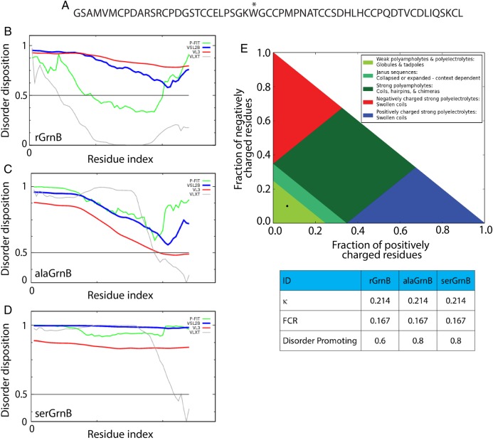
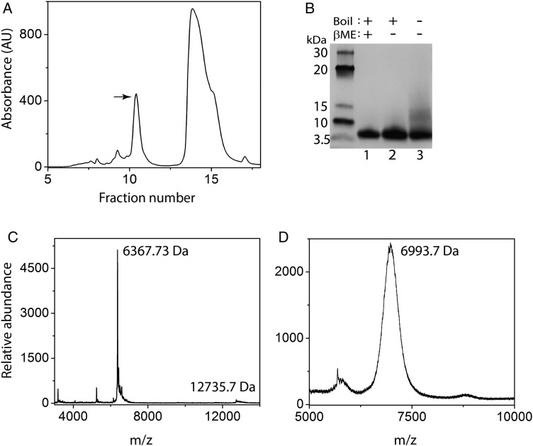
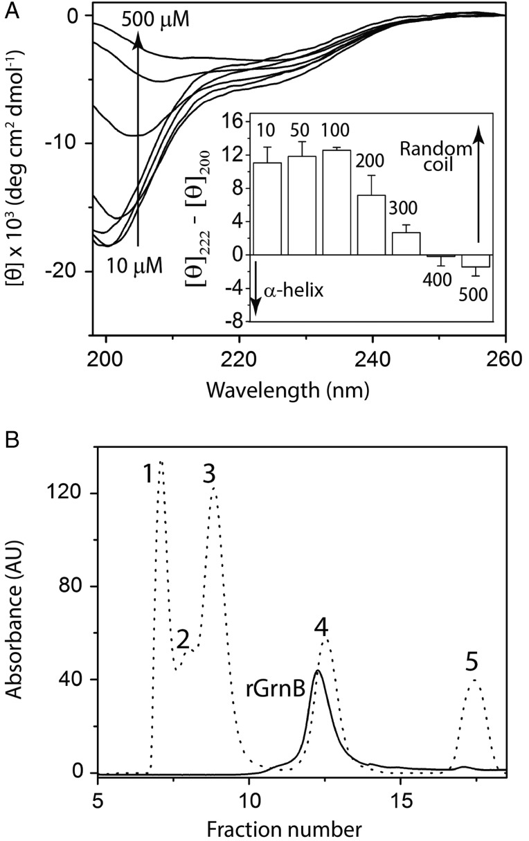
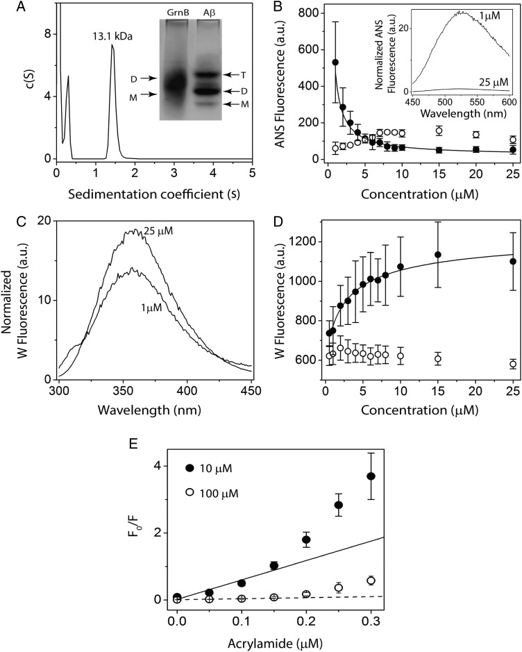
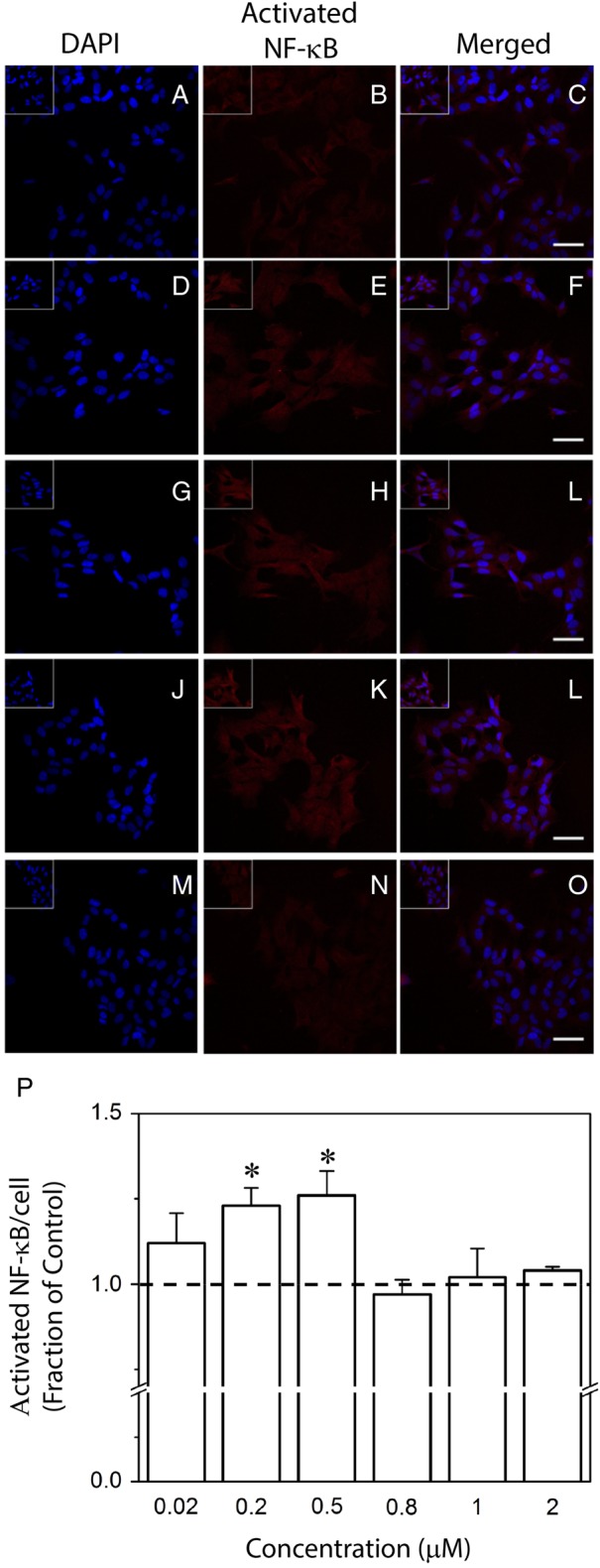
Granulins (Grns) are a family of small, cysteine-rich proteins that are generated upon proteolytic cleavage of their precursor, progranulin (Pgrn). All seven Grns (A-G) contain 12 conserved cysteines that form 6 intramolecular disulfide bonds, rendering this family of proteins unique. Grns are known to play multi-functional roles, including wound healing, embryonic growth, and inflammation and are implicated in neurodegenerative diseases. Despite their manifold functions, there exists a dearth of information regarding their structure-function relationship. Here, we sought to establish the role of disulfide bonds in promoting structure by investigating the fully reduced GrnB (rGrnB). We report that monomeric rGrnB is an intrinsically disordered protein (IDP) at low concentrations. rGrnB undergoes dimerization at higher concentrations to form a fuzzy complex without a net gain in the structure-a behavior increasingly identified as a hallmark of some IDPs. Interestingly, we show that rGrnB is also able to activate NF-κB in human neuroblastoma cells in a concentration-dependent manner. This activation correlates with the observed monomer-dimer dynamics. Collectively, the presented data establish that the intrinsic disorder of rGrnB governs conformational dynamics within the reduced form of the protein, and suggest that the overall structure of Grns could be entirely dictated by disulfide bonds.

Keywords: cysteine-rich protein; fuzzy complex; granulin; intrinsically disordered protein; progranulin.


Figures

Fig. 1
Fig. 1
In silico analysis of intrinsic disorder of rGrnB, alaGrnB and serGrnB. (A) Primary sequence of the fully reduced rGrnB. The * at position 28 (position 24 in the sequence of native GrnB) indicates the Y to W substitution made within the construct. (B–D) predictor of naturally disordered regions (PONDR) analysis of rGrnB, alaGrnB and serGrnB, respectively, using VLXT (light gray), VSL2B (black), VL3 (dark gray) and PONDR-Fit (medium gray) predictors. (E) CIDER analysis of rGrnB, alaGrnB and serGrnB shows that all three of them (black dot) fall in region 1 (lightest gray) of the diagram of states (plot of positively charged residues vs. fraction of negatively charged residues. The table, obtained from the CIDER analysis, shows a low kappa (κ) value of 0.214, low FCR (fraction of charged residues) value of 0.167 and disorder promoting value of 0.6 for rGrnB and 0.8 for alaGrnB and serGrnB.
Fig. 2
Fig. 2
Characterization of recombinant rGrnB. (A), HPLC profile of rGrnB purification after thrombin cleavage. The peak marked with an arrow indicates the fraction corresponding to the native form of rGrnB. The other peak corresponds to thioredoxin. (B) SDS-PAGE analysis of the rGrnB fraction marked with an arrow on the HPLC profile. Lane 1 contains the sample reduced using 2-mercaptoethanol (β-ME) and boiled; Lane 2 contains nonreduced and boiled sample; Lane 3 contains nonreduced and not boiled sample. (C) MALDI-ToF analysis of rGrnB indicating a molecular weight of 6367.39 Da, corresponding to monomeric mass (theoretical MW 6367.4 Da). In addition, the spectrum shows the presence of rGrnB dimer (12735.3 Da). (D) Alkylation of rGrnB with iodoacetamide showing a signal corresponding to 6993.7 Da indicating presence of 10.7 (∼11) free thiols (reduced cysteines). The results are representative of three or more consistent repeats.
Fig. 3
Fig. 3
rGrnB is an IDP. (A) Concentration-dependent conformational changes in rGrnB observed by far-UV CD. (Inset) Spectra as a difference in the molar ellipticities at 222 nm (helix) and 200 nm (random coil). (B) Fractionation of rGrnB using a Superdex™ 75 size exclusion column (solid line) and compared with the fractionation profile of gel filtration standards (dotted line)—1-bovine thyroglobulin (670 kDa), 2-bovine γ-globulin (158 kDa), 3-chicken ovalbumin (44 kDa), 4-horse myoglobin (17 kDa) and 5-vitamin B12 (1.35 kDa).
Fig. 4
Fig. 4
Effect of osmolytes on rGrnB conformation. (A) Far UV CD spectra of rGrnB measured in the presence of increasing concentrations of TFE. (Inset) The difference in the ellipticities between 208 and 222 nm (helix) for each TFE concentration plotted against the concentration of TFE. The data were fitted with Boltzman's sigmoidal equation to obtain a melting concentration of 16 ± 5% TFE. (B) Far UV CD spectra of rGrnB in the presence of increasing concentrations of trimethylamine N-oxide (TMAO).
Fig. 5
Fig. 5
Dimerization of rGrnB. (A) Molecular size distribution of rGrnB at 100 μM obtained by sedimentation velocity indicates a predominant dimer (MW 13.1 kDa). (Inset) Native PAGE analysis of rGrnB at 100 μM run on a 14% gel indicates a diffuse dimeric band corresponding to ∼13 kDa, based on the Aβ sample electrophoresed in parallel for which monomer (M: 4.5 kDa), dimer (D: 9.0 kDa) and trimer (T: 13.5 kDa) are observed. (B, D) rGrnB (closed circle) concentration-dependent ANS binding and intrinsic tryptophan fluorescence, respectively. The normalized fluorescence was plotted against protein concentration, and data were fitted (solid line) to a monomer–dimer model as described in Supplementary data. Bovine serum albumin (BSA) (open circle) was used as a negative control. (Inset) (B) Representative normalized ANS fluorescence scans of 25 and 1 μM rGrnB. (C) Representative normalized tryptophan fluorescence scans of 25 and 1 μM rGrnB. E, Normalized Stern-Volmer plots for 10 μM (closed circle) and 100 μM (open circle) rGrnB using acrylamide as the quenching agent.
Fig. 6
Fig. 6
Effect of rGrnB on NF-κB activation in SH-SY5Y cells. SH-SY5Y cells were incubated alone (control, A–C) or in the presence of 0.02 (D–F), 0.2 (G–I), 0.5 (J–L), 0.8 (not shown), 1 (not shown), or 2 μM (M–O) rGrnB. Immunofluorescence staining was performed for the activated form of NF-κB in conjunction with nuclear DAPI staining. Scale bars = 50 μm. Inserts located in top left corners represent ×1.5 magnification. (P) Activated NF-κB staining intensity per cell was analyzed using a custom MATLAB routine as described in the Experimental procedures. Results are reported relative to the control. Error bars indicate SEM, n = 3–4. *P < 0.05, relative to the control.

References

    1. Abràmoff M.D., Magalhães P.J., Ram S.J. (2004) Biophotonics Int., 11, 36–43.
    1. Baker M., Mackenzie I.R., Pickering-Brown S.M. et al. (2006) Nature, 442, 916–919. - PubMed
    1. Baldwin A.S. (1996) Annu. Rev. Immunol., 14, 649–681. - PubMed
    1. Bateman A., Bennett H.P. (1998) J. Endocrinol., 158, 145–151. - PubMed
    1. Boteva R., Zlateva T., Dorovska-Taran V., Visser A.J., Tsanev R., Salvato B. (1996) Biochemistry, 35, 14825–14830. - PubMed

Publication types

MeSH terms

Substances