Intra-Individual, Inter-Vendor Comparison of Diffusion-Weighted MR Imaging of Upper Abdominal Organs at 3.0 Tesla with an Emphasis on the Value of Normalization with the Spleen
- PMID: 26957905
- PMCID: PMC4781759
- DOI: 10.3348/kjr.2016.17.2.209
Intra-Individual, Inter-Vendor Comparison of Diffusion-Weighted MR Imaging of Upper Abdominal Organs at 3.0 Tesla with an Emphasis on the Value of Normalization with the Spleen
Abstract
Objective: To compare the apparent diffusion coefficient (ADC) values of upper abdominal organs with 2 different 3.0 tesla MR systems and to investigate the usefulness of normalization using the spleen.
Materials and methods: Forty-one patients were enrolled in this prospective study, of which, 35 patients (M:F, 27:8; mean age ± standard deviation, 62.3 ± 12.3 years) were finally analyzed. In addition to the routine liver MR protocol, single-shot spin-echo echo-planar diffusion-weighted imaging using b values of 0, 50, 400, and 800 s/mm(2) in 2 different MR systems was performed. ADC values of the liver, spleen, pancreas, kidney and liver lesion (if present) were measured and analyzed. ADC values of the spleen were used for normalization. The Pearson correlation, Spearman correlation, paired sample t test, Wilcoxon signed rank test and Bland-Altman method were used for statistical analysis.
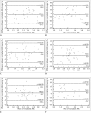
Results: For all anatomical regions and liver lesions, both non-normalized and normalized ADC values from 2 different MR systems showed significant correlations (r = 0.5196-0.8488). Non-normalized ADC values of both MR systems differed significantly in all anatomical regions and liver lesions (p < 0.001). However, the normalized ADC of all anatomical regions and liver lesions did not differ significantly (p = 0.065-0.661), with significantly lower coefficient of variance than that of non-normalized ADC (p < 0.009).
Conclusion: Normalization of the abdominal ADC values using the spleen as a reference organ reduces differences between different MR systems, and could facilitate consistent use of ADC as an imaging biomarker for multi-center or longitudinal studies.
Keywords: 3.0 T; Apparent diffusion coefficient; Diffusion-weighted imaging; Inter-vendor differences.
Figures


References
-
- Thoeny HC, De Keyzer F. Extracranial applications of diffusion-weighted magnetic resonance imaging. Eur Radiol. 2007;17:1385–1393. - PubMed
-
- Koh DM, Takahara T, Imai Y, Collins DJ. Practical aspects of assessing tumors using clinical diffusion-weighted imaging in the body. Magn Reson Med Sci. 2007;6:211–224. - PubMed
-
- Patterson DM, Padhani AR, Collins DJ. Technology insight: water diffusion MRI--a potential new biomarker of response to cancer therapy. Nat Clin Pract Oncol. 2008;5:220–233. - PubMed
-
- Koinuma M, Ohashi I, Hanafusa K, Shibuya H. Apparent diffusion coefficient measurements with diffusion-weighted magnetic resonance imaging for evaluation of hepatic fibrosis. J Magn Reson Imaging. 2005;22:80–85. - PubMed
-
- Lewin M, Poujol-Robert A, Boëlle PY, Wendum D, Lasnier E, Viallon M, et al. Diffusion-weighted magnetic resonance imaging for the assessment of fibrosis in chronic hepatitis C. Hepatology. 2007;46:658–665. - PubMed
MeSH terms
LinkOut - more resources
Full Text Sources
Other Literature Sources

