. 2016 Mar 8:7:6.
doi: 10.1186/s13227-016-0042-x. eCollection 2016.

The ontogeny of choanocyte chambers during metamorphosis in the demosponge Amphimedon queenslandica

Affiliations

The ontogeny of choanocyte chambers during metamorphosis in the demosponge Amphimedon queenslandica

Shunsuke Sogabe et al. Evodevo. .

Abstract

Background: The aquiferous body plan of poriferans revolves around internal chambers comprised of choanocytes, a cell type structurally similar to choanoflagellates. These choanocyte chambers perform a range of physiological and developmental functions, including the capture of food and the generation of stem cells. Despite the increasing interest for choanocytes as sponge stem cells, there is limited knowledge on the development of choanocyte chambers. Using a combination of cell lineage tracing, antibody staining and EdU labeling, here we examine the development of choanocytes and the chambers they comprise during metamorphosis in the marine demosponge Amphimedon queenslandica.

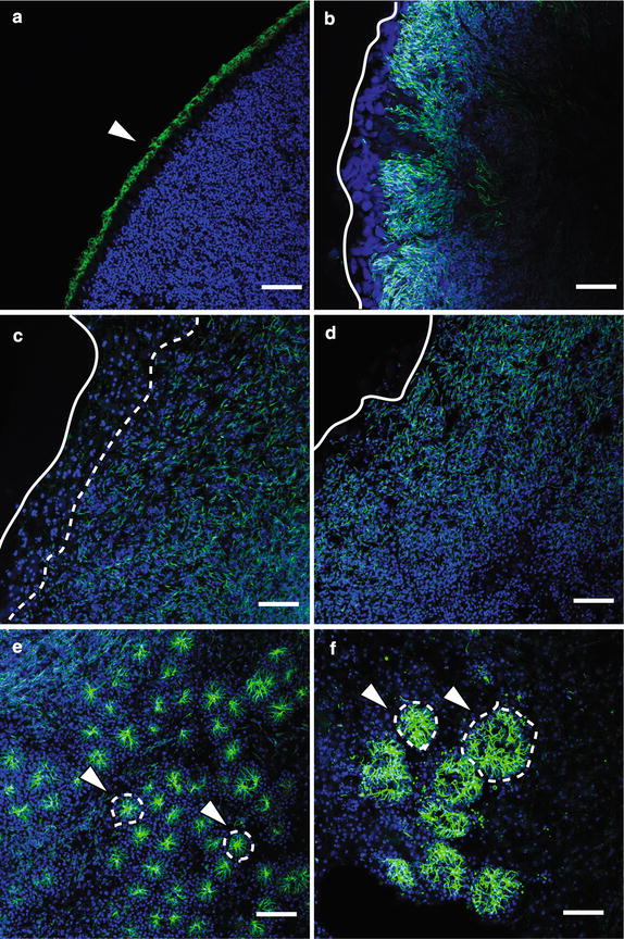
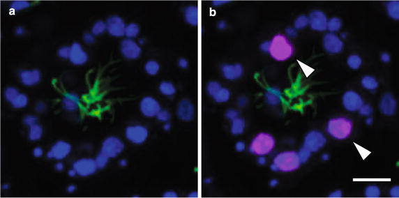
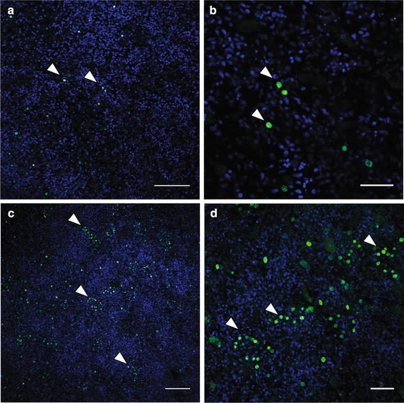
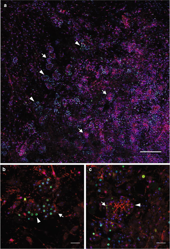
Results: Lineage-tracing experiments show that larval epithelial cells transform into mesenchymal pluripotent stem cells, resembling archeocytes, within 24 h of initiating metamorphosis. By 36 h, some of these labeled archeocyte-like cells have differentiated into choanocytes that will form the first postlarval choanocyte chambers. Non-labeled cells also contribute to these primary choanocyte chambers, consistent with these chambers being a chimera of multiple transdifferentiated larval cell types and not the proliferation of a single choanocyte precursor. Moreover, cell proliferation assays demonstrate that, following the initial formation of choanocyte chambers, chambers grow at least partially by the proliferation of choanocytes within the chamber, although recruitment of individual cells into established chambers also appears to occur. EdU labeling of postlarvae and juveniles reveals that choanocyte chambers are the primary location of cell proliferation during metamorphosis.

Conclusion: Our results show that multiple larval cell lineages typically contribute to formation of individual choanocyte chambers at metamorphosis, contrary to previous reports in other species that show sponge choanocyte chambers form clonally. Choanocytes in postlarval and juvenile A. queenslandica chambers can also divide, with choanocyte chambers being the primary location of cell proliferation. Interestingly, the level of cell proliferation varies greatly between chambers and appears to be contingent on the size, location and developmental state of the chamber. Small chambers on the periphery of the body tend to possess more dividing cells. As choanocytes can also dedifferentiate into archeocyte-like cells, cell proliferation in chambers may not only contribute to chamber growth and self-renewal but also increase the number of pluripotent archeocytes.

Keywords: Archeocyte; Choanocyte; Choanocyte chamber; Demospongiae; Development; Evolution; Invertebrates; Ontogeny; Porifera; Stem cells.


Figures

Fig. 1
Fig. 1
Anatomy of a choanocyte chamber in A. queenslandica. a A schematic drawing of a choanocyte chamber consisting of multiple choanocytes with a collar of microvilli and cilium pointing inward. b A confocal section of a choanocyte chamber in A. queenslandica consisting of multiple choanocytes with cilia pointing inward. Nuclei (nu) are stained with DAPI (blue); cilia (ci) are immunofluorescently labeled with an anti-acetylated alpha tubulin antibody as well as CM-DiI, which also label the cell body (red). ci cilium, mi microvilli, nu nucleus. Scale bar 5 μm
Fig. 2
Fig. 2
Ciliation pattern changes show choanocyte chamber formation timing during metamorphosis in A. queenslandica. Nuclei are stained with DAPI (blue), and cilia are immunofluorescently labeled with an anti-acetylated alpha tubulin antibody (green). a A confocal section of the external epithelium of a competent larva. Cilia occur predominantly on the apical surface of epithelial cells (arrowhead), while there is no evidence of ciliation in the inner cell mass. b 6 hour postresettlement (hpr). The epithelial integrity is lost [28]. Cilia are becoming resorbed into former epithelial cells [36], and thus, ciliation is no longer found on the external surface of the postlarva (white line). c 12 hpr. Overall ciliation is reduced. Cells containing the resorbed cilia are internalized (white line) and have not yet reached the edge of the body (white line). d 24 hpr. Cells containing resorbed cilia spread across the body (white line). e 48 hpr. Small choanocyte chambers are forming throughout the body (arrowheads, circled by white dotted line), with some cells containing resorbed cilia still visible. f 72 hpr. Larger choanocyte chambers are present (arrowheads, circled by white dotted line), with fewer cells containing resorbed cilia visible. Scale bars 25 μm
Fig. 3
Fig. 3
Larval epithelial cells transform into archeocytes during early metamorphosis. Nuclei are stained with DAPI (blue), and subsets of cells are labeled with CM-DiI (red) or phallacidin (green). a A confocal section of a competent larva labeled with CM-DiI. CM-DiI-labeled cells are present only in the epithelial layer of the larva. The dotted line indicates a boundary between the epithelial layer and subepithelial layer with the inner cell mass. b Confocal section of the larval epithelial layer showing ciliated columnar epithelial cells labeled with CM-DiI (arrowhead, circled by dotted line). Globular (spherulous) cells are non-specifically labeled by phallacidin (arrow, circled by dotted line). c Confocal section of archeocytes found during early metamorphosis in 24 hpr postlarvae. d A high-magnification image of an archeocyte. Small DAPI signals (arrows) are putative nuclear fragments obtained via phagocytosis of larval cells that underwent apoptosis at metamorphosis [28, 42]. The arrowhead shows the much larger and less heavily stained nucleus (with a nucleolus) of this archeocyte. Scale bars a 40 μm; bd 15 μm
Fig. 4
Fig. 4
Evidence of non-clonal choanocyte chambers visualized by differential CM-DiI labeling of postlarval and juvenile choanocyte chambers. Nuclei are stained with DAPI (blue), and cells derived from the larval epithelium are labeled with CM-DiI (red). a A choanocyte chamber completely labeled with CM-DiI. This chamber could have formed by a single progenitor cell (clonal) or multiple CM-DiI-labeled progenitor cells (non-clonal). b A non-clonal choanocyte chamber indicated by a large cluster of CM-DiI-labeled (arrowhead) and unlabeled choanocytes in a single chamber. c Two small clusters of CM-DiI-labeled choanocytes (arrowheads) in a largely unlabeled chamber. The arrow shows a neighboring archeocyte also labeled with CM-DiI. (D) Unlabeled choanocyte chambers (circled by dotted line) and a single choanocyte in the extracellular matrix (arrowhead). CM-DiI-labeled vesicles are visible within the unlabeled choanocytes. Scale bars a, b, 5 μm; c, d 10 μm
Fig. 5
Fig. 5
Choanocyte chambers labeled with anti-PH3 labeled nuclei in a 72-hpr juvenile. a, b Confocal sections of single choanocyte chambers. Nuclei are stained with DAPI (blue), and cilia are immunofluorescently labeled with an anti-acetylated alpha tubulin antibody (green), and mitotic nuclei are labeled with anti-phospho-histone H3 antibody (magenta). Note in b that a subset of choanocytes in the chamber is PH3 positive, indicative of mitosis (arrowheads). Scale bar 5 μm
Fig. 6
Fig. 6
Proliferating nuclei labeled with EdU during early postlarval development. Nuclei are stained with DAPI (blue) and mitotic nuclei pulse labeled with EdU (green). a, b 24 hpr postlarva pulse labeled with EdU from 18 to 24 hpr. A small number of nuclei are labeled with EdU (arrowheads). c, d 30 hpr postlarva pulse labeled with EdU (from 24 to 30 hpr). An increase in proliferation rate can be observed, with clusters of EdU-positive nuclei evident as well (arrowheads). Scale bars a, c 40 μm; b, d 15 μm
Fig. 7
Fig. 7
Cells in early choanocyte chambers labeled with EdU. Nuclei are stained with DAPI (blue) and EdU (green), and cilia are immunofluorescently labeled with an anti-acetylated alpha tubulin antibody (red). a A confocal section of a 36 hpr postlarva pulse labeled with EdU from 30 to 36 hpr. Clusters of EdU-positive nuclei are observed. b A high-magnification image of clusters of EdU-positive cells (putative early choanocyte chambers (circled with dotted line) in a 36 hpr postlarva). At this stage, the cilia (arrowheads) are observed inside early choanocyte chambers. Scale bars a 20 μm; b 10 μm
Fig. 8
Fig. 8
EdU labeling in juvenile choanocyte chambers shows variation in proliferation rates of individual chambers. Nuclei are stained with DAPI (blue) and EdU (green), and cilia and cytoplasm immunofluorescently labeled with both anti-acetylated alpha tubulin antibody and anti-tyrosinated alpha-tubulin antibody (red). a Juvenile labeled with EdU from 72 to 78 hpr. The right bottom corner of the micrograph is closer to the center of the juvenile, and the top left corner is the outer edge of the juvenile. Many EdU-positive nuclei are found throughout the juvenile body. There are variations in the number of EdU labeling found in chambers ranging from little to no EdU signals (arrows) to many or all nuclei labeled with EdU (arrowheads). b Choanocyte chambers with numerous EdU labeling. These appear to be early choanocyte chambers with relatively short cilia (arrowhead) or little to no cilia (arrow). c A mature choanocyte chamber with no EdU labeling (arrowhead). The small punctate EdU signals (arrow) are presumably proliferating bacteria. Scale bars a 80 μm; b, c 10 μm
Fig. 9
Fig. 9
Proposed model of choanocyte chamber formation in A. queenslandica. During metamorphosis, postlarval archeocytes form choanocyte chambers from either a single progenitor cell or multiple cells. Choanocyte chambers grow by proliferation of choanocytes within the chamber and by recruitment of choanocytes into mature chambers

References

    1. Simpson TL. The cell biology of sponges. New York: Springer; 1984.
    1. Funayama N. The stem cell system in demosponges: suggested involvement of two types of cells: archeocytes (active stem cells) and choanocytes (food-entrapping flagellated cells) Dev Genes Evol. 2013;223:23–38. doi: 10.1007/s00427-012-0417-5. - DOI - PubMed
    1. Hill MS, Hill AL, Lopez J, Peterson KJ, Pomponi S, Diaz MC, Thacker RW, Adamska M, Boury-Esnault N, Cárdenas P, et al. Reconstruction of family-level phylogenetic relationships within Demospongiae (Porifera) using nuclear encoded housekeeping genes. PLoS ONE. 2013;8:e50437. doi: 10.1371/journal.pone.0050437. - DOI - PMC - PubMed
    1. Whelan NV, Kocot KM, Halanych KM. Employing phylogenomics to resolve the relationships among cnidarians, ctenophores, sponges, placozoans, and bilaterians. Integr Comp Biol. 2015;55:1084–1095. doi: 10.1093/icb/icv037. - DOI - PubMed
    1. Pisani D, Pett W, Dohrmann M, Feuda R, Rota-Stabelli O, Philippe H, Lartillot N, Wörheide G. Genomic data do not support comb jellies as the sister group to all other animals. Proc Natl Acad Sci USA. 2015;112:15402–15407. doi: 10.1073/pnas.1518127112. - DOI - PMC - PubMed

LinkOut - more resources