Dyke-Davidoff-Masson syndrome
- PMID: 26958525
- PMCID: PMC4765277
- DOI: 10.4103/2229-516X.174016
Dyke-Davidoff-Masson syndrome
Abstract
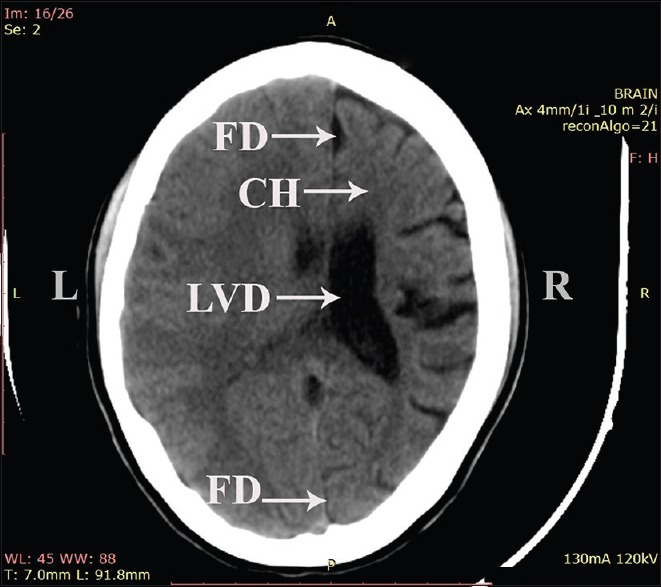
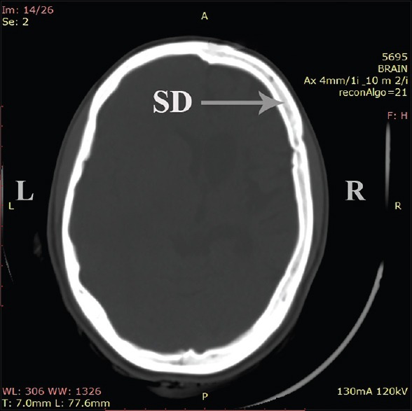
Dyke-Davidoff-Masson syndrome (DDMS) refers to atrophy or hypoplasia of one cerebral hemisphere, due to an insult to the developing brain in fetal or early childhood period. Age of presentation depends on the time of neurologic insult, and characteristic changes may be seen only in adolescence. Male gender and left hemisphere are more frequently involved. A 17-year-old female adolescent with a history of recurrent refractory seizures, hemiplegia and mental retardation reported to Department of Radiology for computed tomography (CT) assessment of brain. On examination, she had facial asymmetry, delayed milestones, and spastic hemiplegia. The CT brain showed right cortical atrophy with ventricular dilatation, prominent sulci, and shifting of falx to the right side. Bone window image showed asymmetry in skull vault thickness, the width of diploic space, the size of paranasal air sinuses and inclination of the petrous ridge between the affected and normal sides. As the above case deviates from the usual presentation of male left sided DDMS, hence the report.
Keywords: Cerebral atrophy; paranasal air sinuses; petrous ridge; prominent sulci.
Figures

References
-
- Dyke CG, Davidoff LM, Masson CB. Cerebral hemiatrophy with homolateral hypertrophy of the skull and sinuses. Surg Gynecol Obstet. 1933;57:588–600.
-
- Aguiar PH, Liu CW, Leitão H, Issa F, Lepski G, Figueiredo EG, et al. MR and CT imaging in the Dyke-Davidoff-Masson syndrome. Report of three cases and contribution to pathogenesis and differential diagnosis. Arq Neuropsiquiatr. 1998;56:803–7. - PubMed
-
- Unal O, Tombul T, Cirak B, Anlar O, Incesu L, Kayan M. Left hemisphere and male sex dominance of cerebral hemiatrophy (Dyke-Davidoff-Masson Syndrome) Clin Imaging. 2004;28:163–5. - PubMed
-
- Shrestha B. Acquired cerebral hemiatrophy: Dyke-Davidoff-Masson Syndrome - A case report. Turk Neurosurg. 2013;23:117–21. - PubMed
-
- Shen WC, Chen CC, Lee SK, Ho YJ, Lee KR. Magnetic resonance imaging of cerebral hemiatrophy. J Formos Med Assoc. 1993;92:995–1000. - PubMed
Publication types
LinkOut - more resources
Full Text Sources
Other Literature Sources
Research Materials