

Review
doi: 10.1016/j.bpj.2016.02.006.
How Viruses Invade Cells
Affiliations
- PMID: 26958878
- PMCID: PMC4788752
- DOI: 10.1016/j.bpj.2016.02.006
Item in Clipboard
Review
How Viruses Invade Cells
Biophys J.
.
No abstract available
Figures

Viral entry pathways. Virus can fuse either directly to the plasma membrane (receptor-mediated fusion) or after being swallowed into an endosome. Which of these routes is followed depends on the type of virus. In fusion with the plasma membrane, the virus binds to a protein in the cell membrane. The function of this cellular protein (a receptor for the virus, shown in green) is perverted to induce a conformational change in the viral fusion protein, leading to fusion. For virus that is triggered within an endosome, the endosome’s acidic conditions induce fusion. In either case, the viral genome passes through a fusion pore into cytosol, and infection is initiated. To see this figure in color, go online.
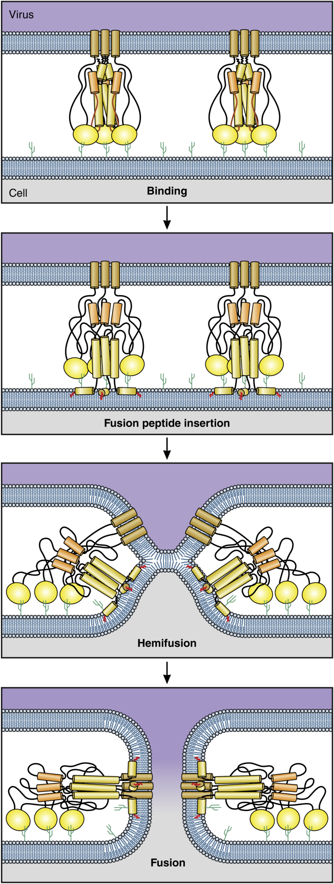
The steps of fusion. Virus binds to specific receptors (each illustrated as a small cactus) on a cell membrane. Initially, four monolayers (in blue) separate the two interior aqueous compartments. After fusion peptides insert into the target membrane, monolayers that face each other merge and clear from the merged region. The noncontacting monolayers bend into the cleared region and come into contact with each other, forming a new bilayer membrane known as a hemifusion diaphragm. At this point (hemifusion), only two monolayers separate the compartments. The fusion protein acts as a nutcracker to force the formation of a pore within the hemifusion diaphragm. This establishes continuity between the two aqueous compartments and fusion is complete. To see this figure in color, go online.
References
-
- Zhu P., Liu J., Roux K.H. Distribution and three-dimensional structure of AIDS virus envelope spikes. Nature. 2006;441:847–852. - PubMed
-
- Cohen F.S., Melikyan G.B. The energetics of membrane fusion from binding, through hemifusion, pore formation, and pore enlargement. J. Membr. Biol. 2004;199:1–14. - PubMed
Publication types
MeSH terms
Grants and funding
LinkOut - more resources
Full Text Sources
Other Literature Sources
