Review
. 2015 Nov;2(1):241-64.
doi: 10.1146/annurev-virology-100114-055043.

Retroviral Integrase: Then and Now

Affiliations
Review

Retroviral Integrase: Then and Now

Mark D Andrake et al. Annu Rev Virol. 2015 Nov.

Abstract

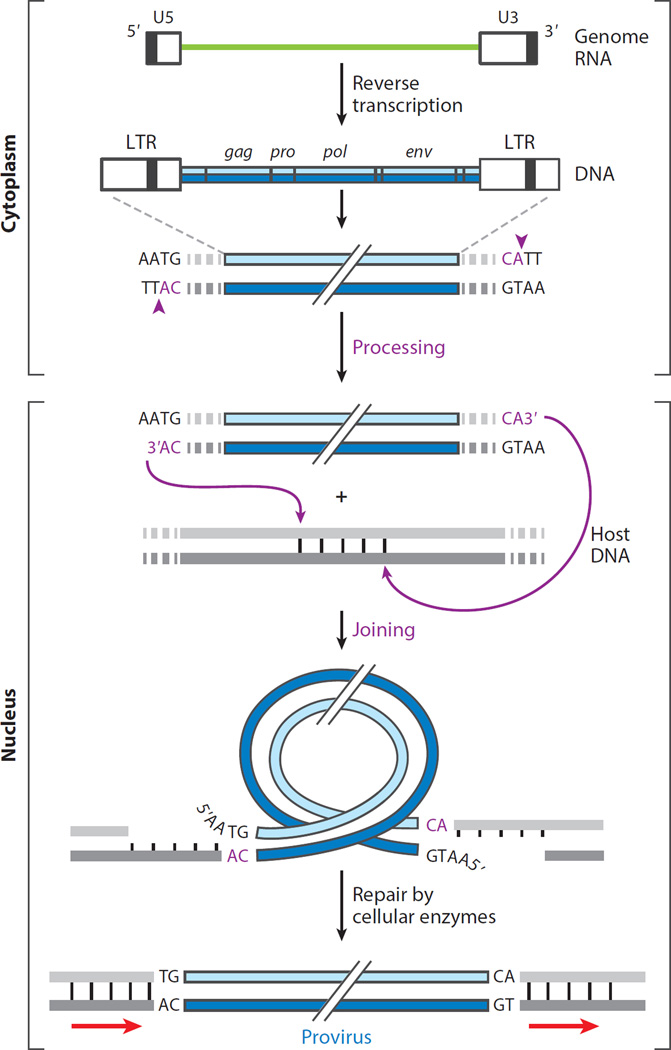
The retroviral integrases are virally encoded, specialized recombinases that catalyze the insertion of viral DNA into the host cell's DNA, a process that is essential for virus propagation. We have learned a great deal since the existence of an integrated form of retroviral DNA (the provirus) was first proposed by Howard Temin in 1964. Initial studies focused on the genetics and biochemistry of avian and murine virus DNA integration, but the pace of discovery increased substantially with advances in technology, and an influx of investigators focused on the human immunodeficiency virus. We begin with a brief account of the scientific landscape in which some of the earliest discoveries were made, and summarize research that led to our current understanding of the biochemistry of integration. A more detailed account of recent analyses of integrase structure follows, as they have provided valuable insights into enzyme function and raised important new questions.

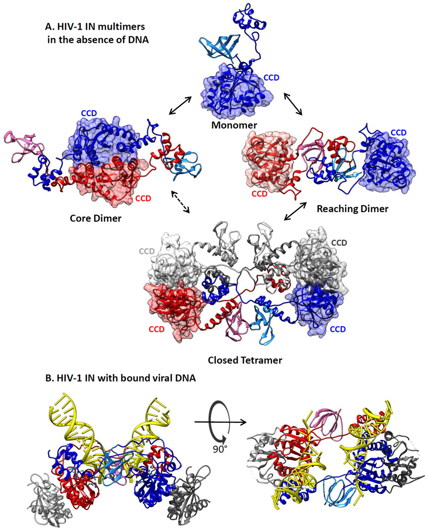
Keywords: core dimer; intasome; integrase; prophage; provirus; reaching dimer.


Figures

Figure 1
Figure 1
Steps in the synthesis of retroviral DNA and its integration into host DNA. The viral RNA genome (green line) is reverse transcribed in the cytoplasm of the cell within a subviral nucleoprotein structure (called the reverse transcription complex) to form a duplex DNA containing long terminal repeats (LTRs) of sequences unique to the 5′ (U5) and 3′ (U3) ends of the viral RNA. The organization of the genes common to all retroviruses (gag, pro, pol, and env) is colinear with the RNA genome. Imperfect inverted repeats at the LTR duplex termini are recognized and nicked by cognate integrase (IN) proteins, following a conserved CA dinucleotide at each 3′ end (vertical arrows in inset), producing recessed 3′-OH ends. This first reaction catalyzed by IN is called processing and takes place within a nucleoprotein assembly called a preintegration complex. A tetramer of IN bound to the processed viral DNA (vDNA) ends enters the nucleus where a joining reaction catalyzed by IN connects the 3′-OH ends of the vDNA to staggered phosphates at a target site in the host DNA. The length of the stagger (4–6 bp) is characteristic of the viral IN protein. The conserved CA dinucleotide and steps catalyzed by IN are highlighted in magenta. Removal of the noncomplementary 5′ nucleotides of vDNA and repair of the gaps in host DNA by host enzymes generate a covalently integrated provirus, shorter by 2 bp on either end and flanked by duplications of the target site (arrows).
Figure 2
Figure 2
Domain organization of integrase (IN) proteins from different retroviruses. (a) Maps for the organization of IN proteins from the alpharetrovirus avian sarcoma/leukosis virus (ASLV), the gammaretrovirus murine leukemia virus (MLV), the lentivirus human immunodeficiency virus type 1 (HIV-1), and the spumavirus prototype foamy virus (PFV). Amino acid numbers delineate the start and end of each domain: the N-terminal extension domain (NED; purple); the N-terminal domain (NTD; red), including the HHCC motif; the catalytic core domain (CCD; blue), including the D,D(35)E motif; and the C-terminal domain (CTD; green). The lengths of linkers that connect the domains are indicated below the lines between domains. Domains for which there is no experimentally determined structure from crystallography are in muted colors. (b) Domain models from crystal structures of the HIV-1 NTD (PDB 1K6Y), CCD (PDB 1BIU), and CTD (PDB 1EX4). The Zn2+ ion in the NTD is shown as an aqua sphere; one of the two Mg2+ ions bound in the active site of the CCD is shown as a green sphere. The conserved Glu residue of the D,D(35)E motif is presumed to chelate the second metal ion together with the first conserved Asp residue.
Figure 3
Figure 3
PFV IN tetramer in complex with two vDNA oligonucleotides and a tDNA. (a) The assembled complex (PDB 4E7K) (; see also supplemental movies in 88) is shown in ribbon representation with the inner subunits in red and blue. Only the CCDs of the outer subunits (gray) are resolved. vDNA oligonucleotides are in orange ribbon ladder representation and the tDNA in yellow and black. The locations of the NEDs and CCDs are indicated. (b) DNA components of the complex portrayed before and after joining, rotated 90° about the y axis from panel a. Processed vDNA ends are shown prior to joining (left) and after joining (right) to the target DNA. (c) The complex shown in panel a is pulled apart to show the positions of all domains in the inner subunits. Interactions between the distal NTD and NED of one inner subunit and the vDNA held in the CCD of the other inner subunit are indicated by arrows. Assembly of the complex is shown in Video 1. Abbreviations: CCD, catalytic core domain; CTD, C-terminal domain; IN, integrase; NED, N-terminal extension domain; NTD, N-terminal domain; PFV, prototype foamy virus; tDNA, target DNA; vDNA, viral DNA.
Figure 4
Figure 4
Models for architectures of full-length human immunodeficiency virus type 1 (HIV-1) apo-integrase (IN) protein in solution. (a) Structures for HIV-1 IN protein in the absence of DNA substrates were derived by HADDOCK data-driven modeling of the HIV-1 IN monomer, dimer, and tetramer in solution, based on small-angle X-ray scattering and protein cross-linking data (93). Catalytic core domains (CCDs) are rendered in muted surface representation to emphasize their locations in the structures. It is not yet known which of these forms is competent for the viral DNA binding that leads to assembly of an HIV-1 intasome. (b) An HIV-1 intasome model (90) with DNA shown in yellow ladder representation. Structures, colored as in Figure 3, are in a ribbon rendering and were generated using Chimera software (133).

References

    1. Wollman E, Wollman E. Les «phases» des bactériophages (facteurs lysogènes) C.R. Soc. Biol. 1937;124:931–934.
    1. Lwoff A. Lysogeny. Bacteriol. Rev. 1953;17:269–337. - PMC - PubMed
    1. Jacob F, Wollman E. Sexuality and the Genetics of Bacteria. New York: Academic; 1961.
    1. Campbell A. Episomes. Adv. Genet. 1961;11:101–145.
    1. Hershey AD, Burgi E, Ingraham L. Cohesion of DNA molecules isolated from phage lambda. PNAS. 1963;49:748–755. - PMC - PubMed

Publication types