Review
. 2015 Nov;2(1):265-88.
doi: 10.1146/annurev-virology-100114-055218.

Continuous and Discontinuous RNA Synthesis in Coronaviruses

Affiliations
Review

Continuous and Discontinuous RNA Synthesis in Coronaviruses

Isabel Sola et al. Annu Rev Virol. 2015 Nov.

Abstract

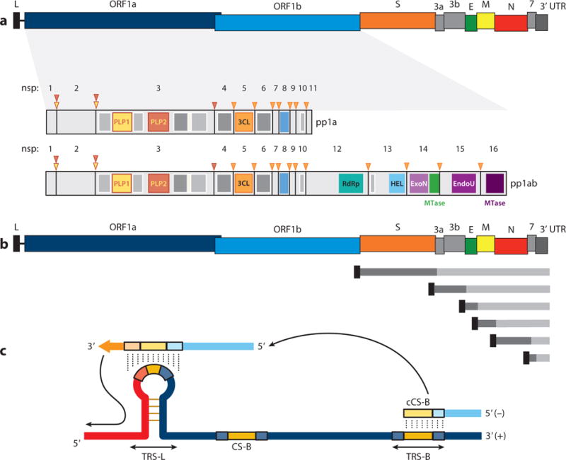
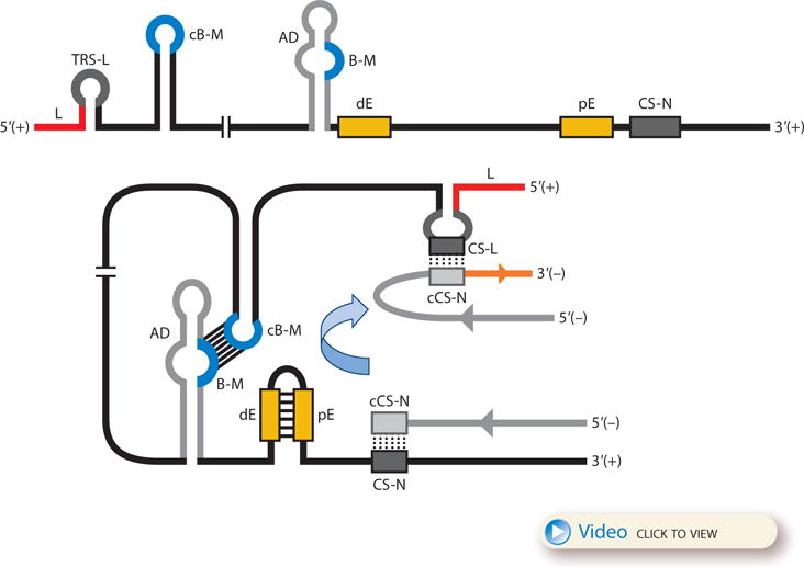
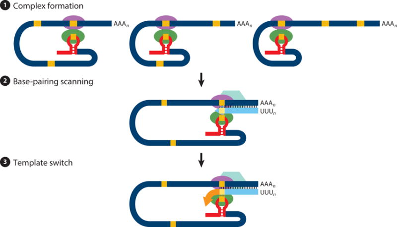
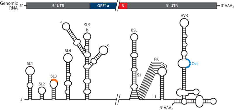
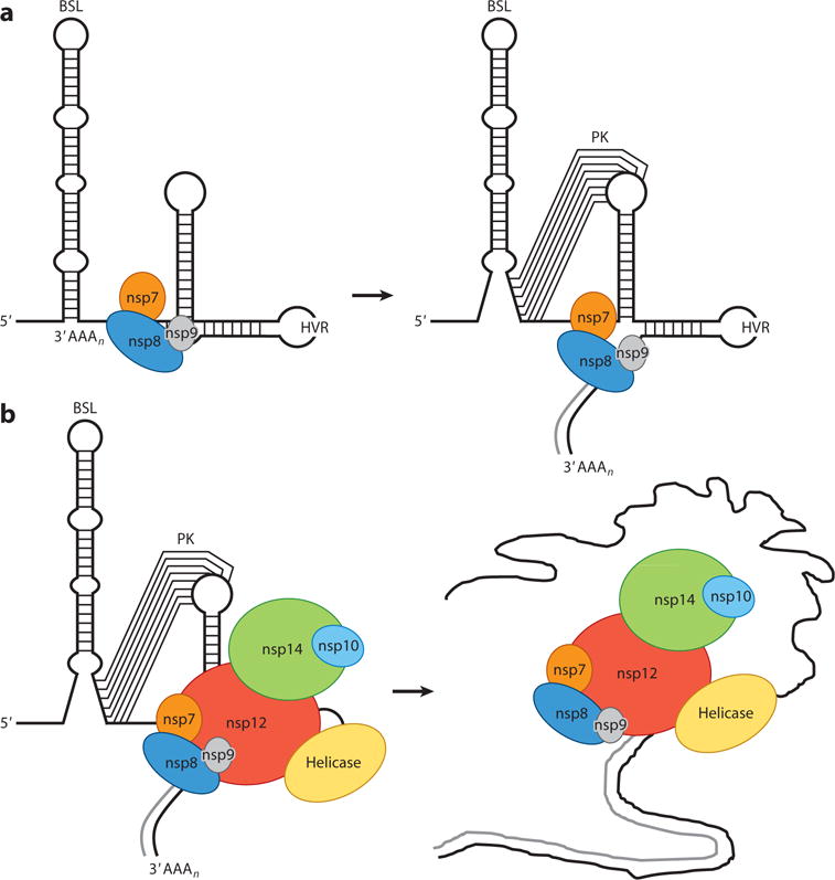
Replication of the coronavirus genome requires continuous RNA synthesis, whereas transcription is a discontinuous process unique among RNA viruses. Transcription includes a template switch during the synthesis of subgenomic negative-strand RNAs to add a copy of the leader sequence. Coronavirus transcription is regulated by multiple factors, including the extent of base-pairing between transcription-regulating sequences of positive and negative polarity, viral and cell protein-RNA binding, and high-order RNA-RNA interactions. Coronavirus RNA synthesis is performed by a replication-transcription complex that includes viral and cell proteins that recognize cis-acting RNA elements mainly located in the highly structured 5' and 3' untranslated regions. In addition to many viral nonstructural proteins, the presence of cell nuclear proteins and the viral nucleocapsid protein increases virus amplification efficacy. Coronavirus RNA synthesis is connected with the formation of double-membrane vesicles and convoluted membranes. Coronaviruses encode proofreading machinery, unique in the RNA virus world, to ensure the maintenance of their large genome size.

Keywords: RNA proofreading; nidovirus; positive-strand RNA viruses; replication; transcription; virus-host interaction.


Conflict of interest statement

DISCLOSURE STATEMENT

The authors are not aware of any affiliations, memberships, funding, or financial holdings that might be perceived as affecting the objectivity of this review.

Figures

Figure 1
Figure 1
Coronavirus genome structure and gene expression. (a) Coronavirus genome structure. The upper scheme represents the TGEV genome. Labels indicate gene names; L corresponds to the leader sequence. Also represented are the nsps derived from processing of the pp1a and pp1ab polyproteins. PLP1, PLP2, and 3CL protease sites are depicted as inverted triangles with the corresponding color code of each protease. Dark gray rectangles represent transmembrane domains, and light gray rectangles indicate other functional domains. (b) Coronavirus genome strategy of sgmRNA expression. The upper scheme represents the TGEV genome. Short lines represent the nested set of sgmRNAs, each containing a common leader sequence (black) and a specific gene to be translated (dark gray). (c) Key elements in coronavirus transcription. A TRS precedes each gene (TRS-B) and includes the core sequence (CS-B) and variable 5′ and 3′ flanking sequences. The TRS of the leader (TRS-L), containing the core sequence (CS-L), is present at the 5′ end of the genome, in an exposed location (orange box in the TRS-L loop). Discontinuous transcription occurs during the synthesis of the negative-strand RNA (light blue), when the copy of the TRS-B hybridizes with the TRS-L. Dotted lines indicate the complementarity between positive-strand and negative-strand RNA sequences. Abbreviations: EndoU, endonuclease; ExoN, exonuclease; HEL, helicase; MTase, methyltransferase (green, N7-methyltransferase; dark purple, 2′-O-methyltransferase); nsp, nonstructural protein; PLP, papain-like protease; RdRp, RNA-dependent RNA polymerase; sgmRNA, subgenomic RNA; TGEV, transmissible gastroenteritis virus; TRS, transcription-regulating sequence; UTR, untranslated region.
Figure 2
Figure 2
Model for the formation of genome high-order structures regulating N gene transcription. The upper linear scheme represents the coronavirus genome. The red line indicates the leader sequence in the 5′ end of the genome. The hairpin indicates the TRS-L. The gray line with arrowheads represents the nascent negative-sense RNA. The curved blue arrow indicates the template switch to the leader sequence during discontinuous transcription. The orange line represents the copy of the leader added to the nascent RNA after the template switch. The RNA-RNA interactions between the pE (nucleotides 26894 to 26903) and dE (nucleotides 26454 to 26463) and between the B-M in the active domain (nucleotides 26412 to 26421) and the cB-M in the 5′ end of the genome (nucleotides 477 to 486) are represented by solid lines. Dotted lines indicate the complementarity between positive-strand and negative-strand RNA sequences. Abbreviations: AD, active domain secondary structure prediction; B-M, B motif; cB-M, complementary copy of the B-M; cCS-N, complementary copy of the CS-N; CS-L, conserved core sequence of the leader; CS-N, conserved core sequence of the N gene; dE, distal element; pE, proximal element; TRS-L, transcription-regulating sequence of the leader. For an animated version of the model, see Video 1 or download a PowerPoint slideshow.
Figure 3
Figure 3
Three-step model of coronavirus transcription. (❶) Complex formation. Proteins binding transcription-regulating sequences are represented by ellipsoids, the leader sequence is indicated with a red bar, and core sequences are indicated with orange boxes. (❷) Base-pairing scanning. Negative-strand RNA is shown in light blue; the transcription complex is represented by a hexagon. Vertical lines indicate complementarity between the genomic RNA and the nascent negative strand. (❸) Template switch. Due to the complementarity between the newly synthesized negative-strand RNA and the transcription-regulating sequence of the leader, template switch to the leader is made by the transcription complex to complete the copy of the leader sequence.
Figure 4
Figure 4
Coronavirus cis-acting RNA elements. The higher-order RNA structures indicated in the diagram are mainly based on studies done in betacoronaviruses. The core sequence within the leader transcription-regulating sequence is shown as an orange box on the top of SL3. Abbreviations: BSL, bulged stem loop; HVR, hypervariable region; L1, loop 1 of the pseudoknot; N, nucleocapsid; Oct, conserved octanucleotide; PK, pseudoknot; S1, stem 1 of the pseudoknot; SL, stem loop; UTR, untranslated region.
Figure 5
Figure 5
Coronavirus replication-transcription complex. (a) After binding of the nsp8, nsp7, and nsp9 complex to the genomic RNA 3′ end, the nsp8 primase activity initiates RNA synthesis de novo. This leads to a conformational change in the 3′-end RNA structure, allowing transition from a BSL to a PK folding. (b) PK formation allows binding of the RNA-dependent RNA polymerase (nsp12) complex, including helicase and nsp14-nsp10. This core replication-transcription complex includes polymerase activity (nsp12 and nsp8), processivity factors (nsp7 and nsp8), and proofreading activity (nsp14 and nsp10). Abbreviations: BSL, bulged stem loop; HVR, hypervariable region; nsp, nonstructural protein; PK, pseudoknot.

References

LITERATURE CITED

    1. de Groot RJ, Baker SC, Baric R, Enjuanes L, Gorbalenya AE, et al. Coronaviridae. In: King AMQ, Adams MJ, Carstens EB, Lefkowitz EJ, editors. Virus Taxonomy: Ninth Report of the International Committee on Taxonomy of Viruses. San Diego, CA: Elsevier Academic; 2012. pp. 774–96.
    1. Coleman CM, Frieman MB. Coronaviruses: important emerging human pathogens. J Virol. 2014;88:5209–12. - PMC - PubMed
    1. Marthaler D, Raymond L, Jiang Y, Collins J, Rossow K, Rovira A. Rapid detection, complete genome sequencing, and phylogenetic analysis of porcine deltacoronavirus. Emerg Infect Dis. 2014;20:1347–50. - PMC - PubMed
    1. Huang YW, Dickerman AW, Pineyro P, Li L, Fang L, et al. Origin, evolution, and genotyping of emergent porcine epidemic diarrhea virus strains in the United States. mBio. 2013;4:e00737–13. - PMC - PubMed
    1. Masters PS. The molecular biology of coronaviruses. Adv Virus Res. 2006;66:193–292. - PMC - PubMed

RELATED RESOURCES

    1. CDC (Cent. Dis. Control Prev.) Coronavirus. Atlanta, GA: CDC; 2015. http://www.cdc.gov/coronavirus/
    1. Enjuanes L, Sola I, Zuñiga S, Moreno JL. Coronavirus RNA synthesis: transcription. In: Thiel V, editor. Coronaviruses: Molecular and Cellular Biology. Norfolk, UK: Caister Academic; 2007. pp. 81–107.
    1. Graham R, Donalson EF, Baric RS. A decade after SARS: strategies for controlling emerging coronaviruses. Nat Rev Microbiol. 2013;11:836–48. - PMC - PubMed
    1. Gralinski LE, Baric RS. Molecular pathology of emerging coronavirus infections. J Pathol. 2015;235:185–95. - PMC - PubMed
    1. Lauber C, Goeman JJ, del Carmen Parquet M, Thi Nga P, Snijder EJ, et al. The footprint of genome architecture in the largest genome expansion in RNA viruses. PLOS Pathog. 2013;9:e1003500. - PMC - PubMed

Publication types