Review
. 2015 Nov;2(1):549-72.
doi: 10.1146/annurev-virology-100114-055249. Epub 2015 Sep 25.

No Love Lost Between Viruses and Interferons

Affiliations
Review

No Love Lost Between Viruses and Interferons

Volker Fensterl et al. Annu Rev Virol. 2015 Nov.

Abstract

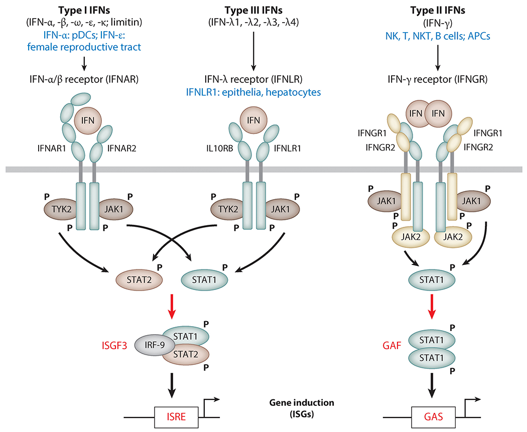
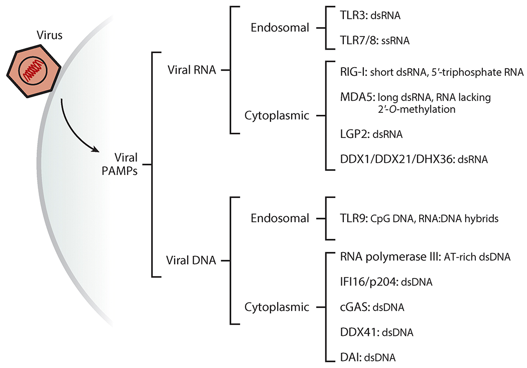
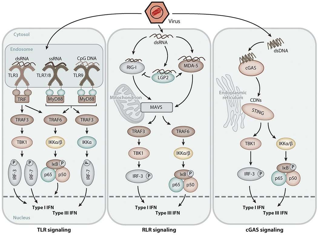
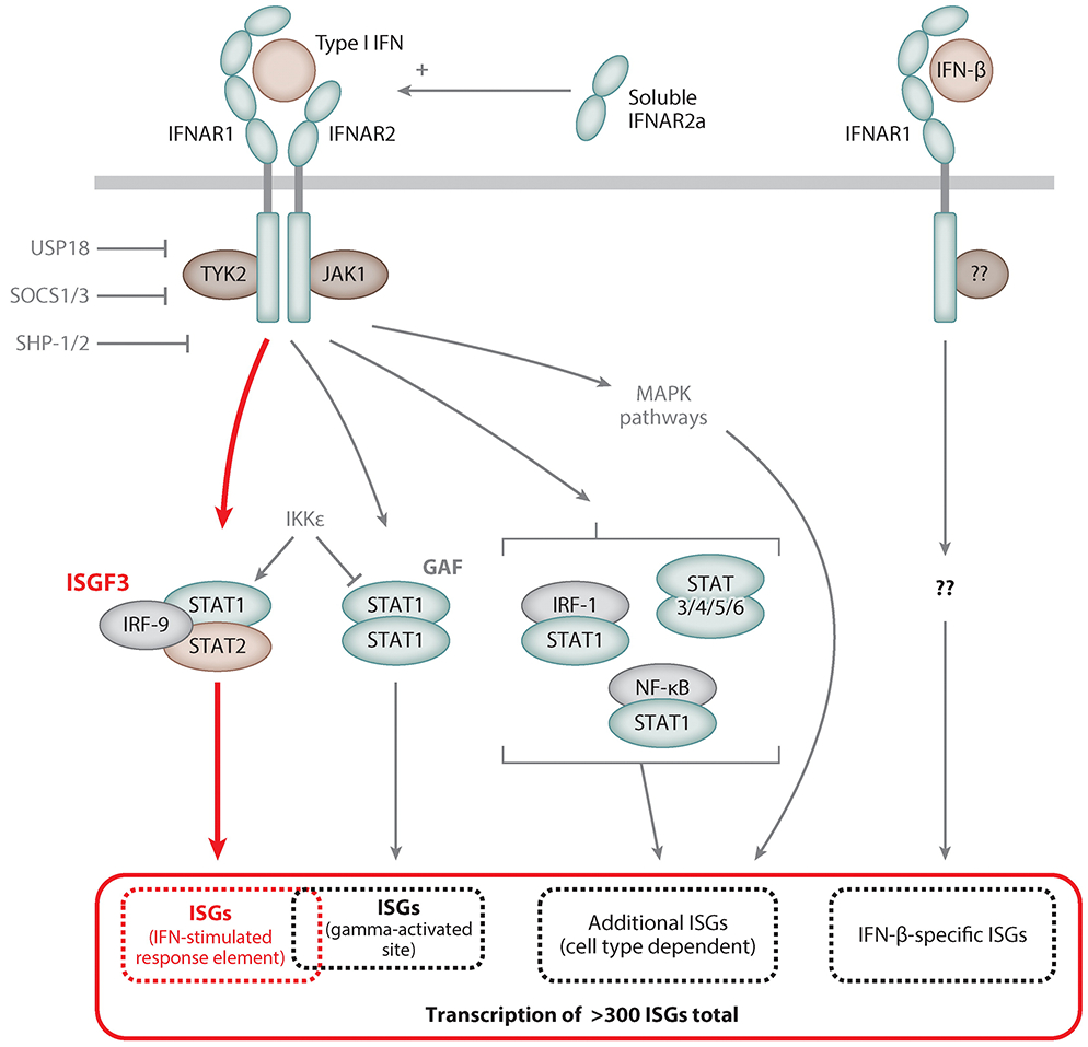
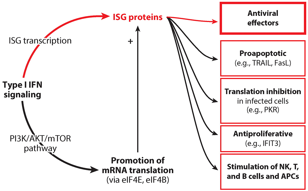
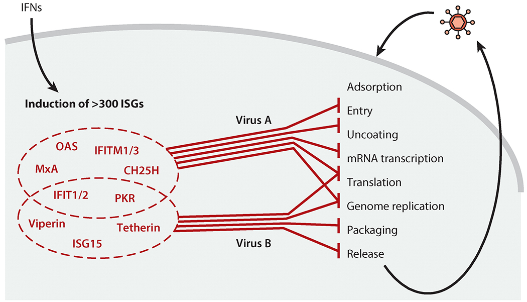
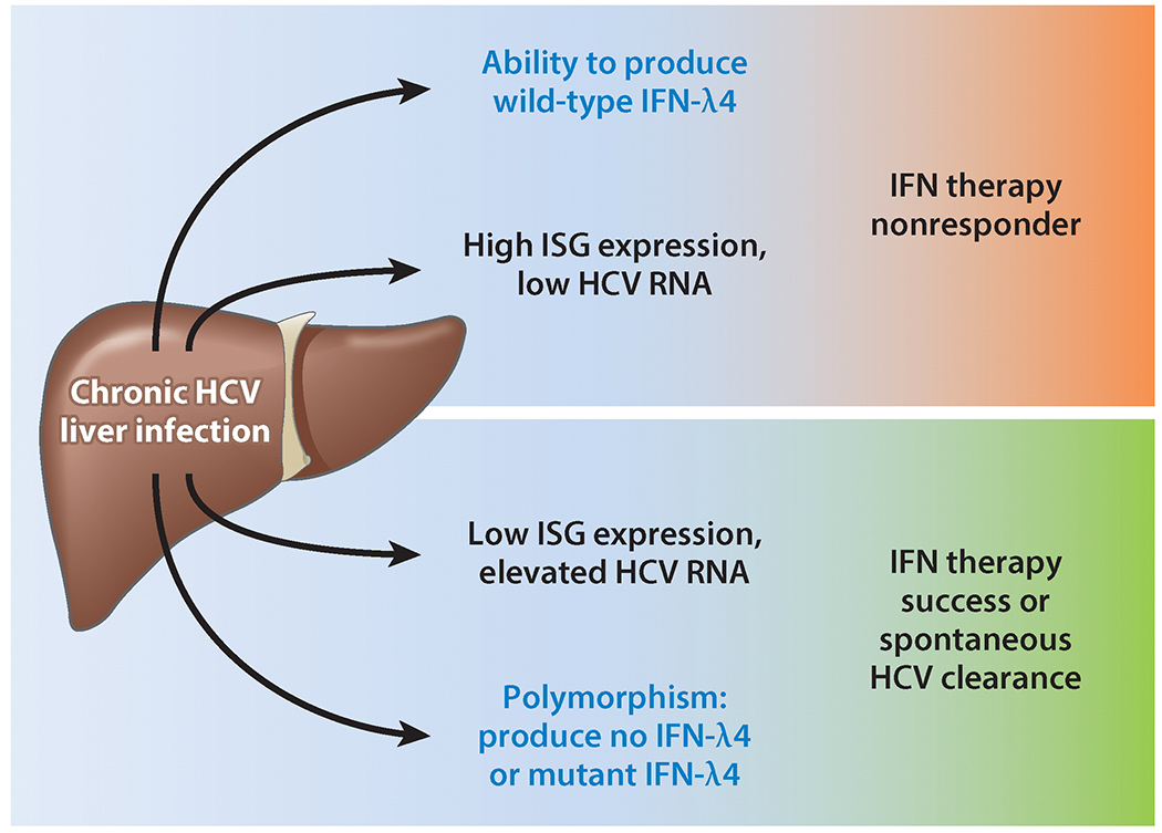
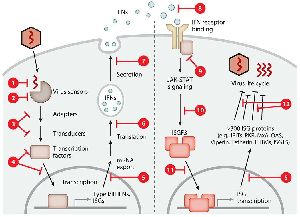
The interferon system protects mammals against virus infections. There are several types of interferons, which are characterized by their ability to inhibit virus replication and resultant pathogenesis by triggering both innate and cell-mediated immune responses. Virus infection is sensed by a variety of cellular pattern-recognition receptors and triggers the synthesis of interferons, which are secreted by the infected cells. In uninfected cells, cell surface receptors recognize the secreted interferons and activate intracellular signaling pathways that induce the expression of interferon-stimulated genes; the proteins encoded by these genes inhibit different stages of virus replication. To avoid extinction, almost all viruses have evolved mechanisms to defend themselves against the interferon system. Consequently, a dynamic equilibrium of survival is established between the virus and its host, an equilibrium that can be shifted to the host's favor by the use of exogenous interferon as a therapeutic antiviral agent.

Keywords: antiviral action; dsRNA; innate immunity; interferon-stimulated gene; interferon-λ; pathogenesis; pattern-recognition receptor; viral evasion; virus infection.


Figures

Figure 1
Figure 1
Major differences between the type I, II, and III IFN systems. Each type of IFN acts through its own cognate cell surface receptor. The Janus kinases JAK1, TYK2, and JAK2 phosphorylate STAT proteins and trigger their transcriptional activity. The three signaling systems are distinguished by their cell type–specific expression of IFNs or their receptor (blue), as well as by the different transcription factors they activate (red). Abbreviations: APC, antigen-presenting cell; GAS, gamma-activated site; IFN, interferon; ISG, interferon-stimulated gene; ISRE, IFN-stimulated response element; NK, natural killer; NKT, natural killer T; pDC, plasmacytoid dendritic cell. To download a PowerPoint slideshow illustrating further details about each IFN system, click the Interactive Figure button.
Figure 2
Figure 2
Cellular sensors detecting viral RNA and DNA. Viral nucleic acids are the key pathogen-associated molecular pattern (PAMP) for cells to recognize the presence of viruses. A large number of sensor proteins (pattern-recognition receptors) bind specific forms of nucleic acids in endosomes and in the cytoplasm and trigger signaling to elicit innate immune responses such as the induction of interferons.
Figure 3
Figure 3
Signaling pathways for type I and III interferon (IFN) induction triggered by viral nucleic acids. Upon virus entry, viral RNA or DNA in endosomes or the cytoplasm is detected by receptor proteins [e.g., Toll-like receptors (TLRs), RIG-I-like receptors (RLRs), cyclic GMP-AMP synthase (cGAS)], which trigger signaling via adapter proteins such as TRIF, MyD88, MAVS, and STING to activate transcription factors of the IRF family as well as NF-κB, leading to induced transcription of the type I and type III IFN genes.
Figure 4
Figure 4
Type I interferon (IFN)-stimulated signaling pathways. Binding of type I IFNs to the dimeric type I IFN receptor, IFNAR, triggers activation of intracellular tyrosine kinases such as JAK1, TYK2, and MAPKs. These in turn activate transcription factor complexes containing STAT proteins, predominantly ISGF3, and others, depending on the cell type. MAPKs activate transcription factors of the ATF/JUN families. IKKϵ serine phosphorylates STAT1 and inhibits its dimerization in favor of ISGF3 formation. A unique interaction of IFN-β with IFNAR1 induces an additional set of genes not induced by other type I IFNs. The soluble splice variant IFNAR2a can enhance type I IFN signaling. Negative regulation of IFNAR signaling is provided by the IFN-stimulated gene (ISG) products USP18 and SOCS, which bind to IFNAR2 and TYK2, respectively, and the tyrosine phosphatases SHP-1 and SHP-2.
Figure 5
Figure 5
Interferon (IFN)-stimulated genes (ISGs): the mediators of the biological effects of IFNs. Type I IFN signaling transcriptionally induces ISGs, whose protein products are the effectors of IFN actions that inhibit virus life cycles. Other ISG proteins mediate proapoptotic effects or general inhibition of translation. Certain cell types show slowed proliferation after continuous IFN exposure. IFN action on immune cells, such as natural killer (NK) cells, T cells, B cells, and antigen-presenting cells (APCs), is important for shaping the innate and adaptive immune responses extrinsic to the infected cells. A transcription-independent effect of IFNs is the activation of mTOR kinase via the AKT pathway, a major promoter of translation.
Figure 6
Figure 6
Inhibition of specific viruses by specific combinations of interferon (IFN)-stimulated genes (ISGs). IFNs induce the expression of hundreds of ISG proteins. Upon subsequent virus infection, a specific subset of ISG proteins inhibits multiple steps of the specific virus’s life cycle; each mechanism contributes to the overall inhibition. Additionally, some ISG proteins are inhibitory only in specific cell types. To download a PowerPoint slideshow illustrating further details about this process, click the Interactive Figure button.
Figure 7
Figure 7
Interferon (IFN)-λ4 as a predictor of responsiveness to IFN-α therapy of chronic hepatitis C virus (HCV) infection. During chronic HCV infection, expression of IFN-λ4 or relatively high IFN-stimulated gene (ISG) expression levels in the liver before treatment correlates with a lack of response to IFN-α2 therapy (i.e., HCV cannot be cleared permanently from the liver). Inversely, being unable to produce IFN-λ4 or having low ISG expression levels makes spontaneous HCV clearance or successful therapy likely. Whether IFN-λ4 is responsible for elevated ISG expression levels is unknown.
Figure 8
Figure 8
Principles of viral antagonism of the interferon (IFN) system. All mammalian viruses evolved to counteract one or multiple phases of the IFN system. The synthesis of IFNs is prevented by viruses by masking their nucleic acid (step ①), inactivation or cleavage of cellular recognition receptors or their adapter proteins (steps ② and ③), inhibition or degradation of key transcription factors such as IRF-3 (step ④), and generalized inhibition of host gene transcription, protein translation, or protein secretion (steps ⑤, ⑥, and ⑦). IFN signaling is blocked by viral expression of soluble decoy receptors (step ⑧), inhibition of Janus kinases (step ⑨), STAT protein sequestration or degradation (step ⑩), or prevention of transcription factor nuclear translocation (step ⑪). Lastly, the actions of antiviral interferon-stimulated gene (ISG) proteins are counteracted by direct antagonism or evasion (step ⑫). To download a PowerPoint slideshow illustrating further details about each step, click the Interactive Figure button.

References

    1. Isaacs A, Lindenmann J. 1957. Virus interference. I. The interferon. Proc. R. Soc. B 147:258–67 - PubMed
    1. Schneider WM, Chevillotte MD, Rice CM. 2014. Interferon-stimulated genes: a complex web of host defenses. Annu. Rev. Immunol 32:513–45 - PMC - PubMed
    1. Versteeg GA, Garcia-Sastre A. 2010. Viral tricks to grid-lock the type I interferon system. Curr. Opin. Microbiol 13:508–16 - PMC - PubMed
    1. McNab F, Mayer-Barber K, Sher A, Wack A, O’Garra A. 2015. Type I interferons in infectious disease. Nat. Rev. Immunol 15:87–103 - PMC - PubMed
    1. de Weerd NA, Nguyen T. 2012. The interferons and their receptors—distribution and regulation. Immunol. Cell Biol 90:483–91 - PMC - PubMed

Publication types