. 2016 Mar 9;11(3):e0150284.
doi: 10.1371/journal.pone.0150284. eCollection 2016.

Chemical and Hormonal Effects on STAT5b-Dependent Sexual Dimorphism of the Liver Transcriptome

Affiliations

Chemical and Hormonal Effects on STAT5b-Dependent Sexual Dimorphism of the Liver Transcriptome

Keiyu Oshida et al. PLoS One. .

Erratum in

Abstract

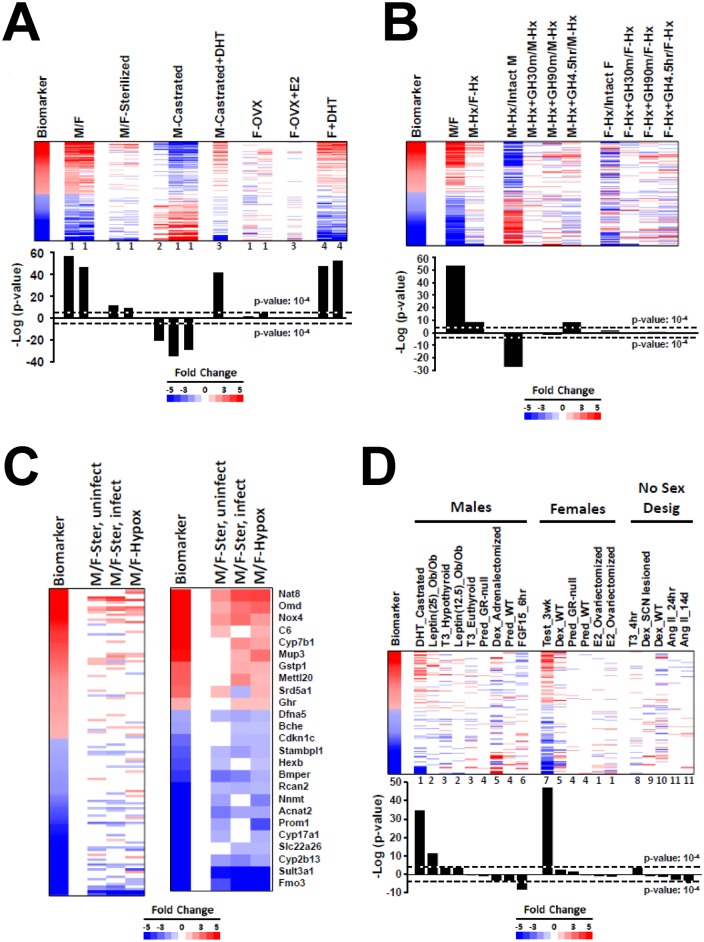
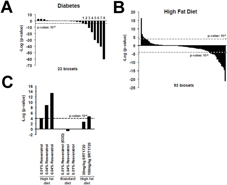
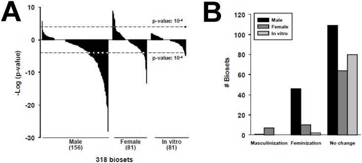
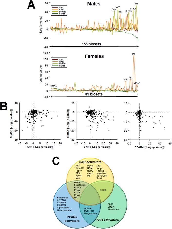
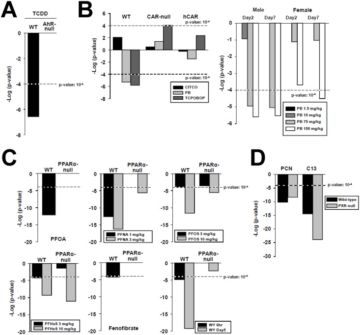
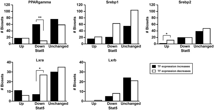
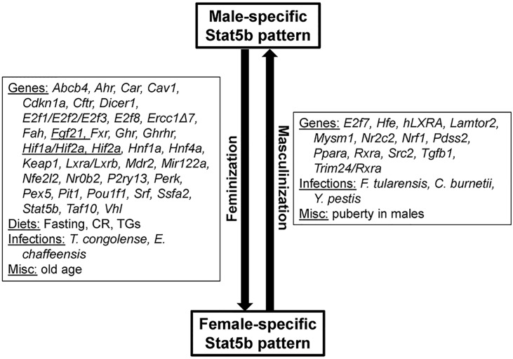
The growth hormone (GH)-activated transcription factor signal transducer and activator of transcription 5b (STAT5b) is a key regulator of sexually dimorphic gene expression in the liver. Suppression of hepatic STAT5b signaling is associated with lipid metabolic dysfunction leading to steatosis and liver cancer. In the companion publication, a STAT5b biomarker gene set was identified and used in a rank-based test to predict both increases and decreases in liver STAT5b activation status/function with high (≥ 97%) accuracy. Here, this computational approach was used to identify chemicals and hormones that activate (masculinize) or suppress (feminize) STAT5b function in a large, annotated mouse liver and primary hepatocyte gene expression compendium. Exposure to dihydrotestosterone and thyroid hormone caused liver masculinization, whereas glucocorticoids, fibroblast growth factor 15, and angiotensin II caused liver feminization. In mouse models of diabetes and obesity, liver feminization was consistently observed and was at least partially reversed by leptin or resveratrol exposure. Chemical-induced feminization of male mouse liver gene expression profiles was a relatively frequent phenomenon: of 156 gene expression biosets from chemically-treated male mice, 29% showed feminization of liver STAT5b function, while <1% showed masculinization. Most (93%) of the biosets that exhibited feminization of male liver were also associated with activation of one or more xenobiotic-responsive receptors, most commonly constitutive activated receptor (CAR) or peroxisome proliferator-activated receptor alpha (PPARα). Feminization was consistently associated with increased expression of peroxisome proliferator-activated receptor gamma (Pparg) but not other lipogenic transcription factors linked to steatosis. GH-activated STAT5b signaling in mouse liver is thus commonly altered by diverse chemicals, and provides a linkage between chemical exposure and dysregulated gene expression associated with adverse effects on the liver.


Conflict of interest statement

Competing Interests: The authors have declared that no competing interests exist.

Figures

Fig 1
Fig 1. Hormonal modulation of liver STAT5b function.
A. Activation of STAT5b function in testosterone-treated mouse liver. The STAT5b biomarker gene set was examined in the indicated biosets from GSE6632, GSE21065, and GSE13265. (Top) Expression behavior of the STAT5b biomarker gene set. The biomarker genes were rank-ordered based on fold-change. Numbers at the bottom of each lane refer to the following studies from which they were derived: 1, GSE21065; 2, GSE6632; 3, GSE13265; 4, GSE13388. (Bottom)–log(p-values) of the similarity to the STAT5b biomarker. M/F, male vs. female; M/F-sterilized, male vs. female comparison in untreated gonadectomized mice; M-castrated, castrated vs. intact mice; M-castrated+DHT, castrated mice treated with dihydrotestosterone (DHT; subcutaneous 5-mg DHT pellet, designed to be released over 21d and giving a plasma level of 1–2 ng DHT/ml vs. castrated mice treated with vehicle; F-OVX, ovariectomized vs. intact mice; F-OVX+E2, ovariectomized estradiol-treated (subcutaneous 0.5-mg E2 pellet (plasma level of 300 pg/ml) designed to be released over 21d) vs. ovariectomized vehicle-treated mice; F+DHT, intact female mice treated with DHT (subcutaneous injections of 100ul sesame oil containing 0.9mg testosterone twice a week for 3 weeks) vs. intact female mice treated with vehicle. B. Effects of pituitary GH modulation on liver STAT5b function. Biosets were derived from Wauthier et al. (2010) (GSE17644) and compared to the STAT5b biomarker gene set. Hypophysectomized (Hypox) male and female mice were given a single ip injection of rat GH (125 ng/g body weight) and killed 30 or 90 min later. Other Hypox male and Hypox female mice were given two ip injections of GH, spaced 4 h apart, and killed 30 min after the second GH injection. (Top) Expression behavior of the STAT5b biomarker genes. Genes were rank-ordered based on fold-change in the biomarker. (Bottom)–log(p-values) of the similarity to the STAT5b biomarker gene set. M/F, male vs female; M-Hx/F-Hx, Hypox males vs. Hypox females; M-Hx/Intact M, Hypox male vs. intact male; M-Hx+GH30m/M-Hx, Hypox males 30 min after a pulse of GH vs. Hypox males; M-Hx+GH90m/M-Hx, Hypox males 90 min after a pulse of GH vs. Hypox males; M-Hx+GH4.5h/M-Hx, Hypox males 4.5 hr after initial exposure to GH vs. Hypox males; F-Hx/Intact F, Hypox female vs. intact female; F-Hx+GH30m/F-Hx, Hypox females 30 min after a pulse of GH vs. Hypox females; F-Hx+GH90m/F-Hx, Hypox females 90 min after a pulse of GH vs. Hypox females; F-Hx+GH4.5h/F-Hx, Hypox females 4.5 hr after initial exposure to GH vs. Hypox females. C. Residual sex differences after sterilization and hypophysectomy. Two biosets from sterilized mice (one from uninfected mice and one from mice infected with Coxiella burnetii, GSE21065) were compared to a bioset from Hypox males vs. Hypox females (GSE17644 described above). (Left) Expression of all genes in the STAT5b biomarker gene set. (Right) Expression of genes that exhibited significant alteration in one or both of the M/F-sterilized biosets and the M-Hx/F-Hx bioset. D. Effects of hormone treatment on liver STAT5b function. Biosets derived from mice treated with the indicated hormones were separated into studies performed in male mice, female mice, and mice with no designation of sex. Numbers at the bottom of each lane refer to the specific studies from which each was derived: 1, GSE13265; 2, GSE19185; 3, GSE32444; 4, GSE21048; 5, GSE24256; 6, GSE29426; 7, GSE13388; 8, GSE21307; 9, GSE564; 10, GSE9630; 11, E-TIGR-12.
Fig 2
Fig 2. Feminization of the liver in models of obesity and diabetes and “re-masculinization” by resveratrol.
A. Feminization in models of diabetes. The biosets which caused feminization (suppression of STAT5b function) were from the following studies: 1) GSE30140; 2) GSE30140; 3) GSE10785; 4) GSE30140; 5) GSE10785; 6) GSE38067; 7) GSE10785; 8) GSE10785. B. Effects of high fat diets on STAT5b. The effects of high fat diet on STAT5b function are based on an analysis of 93 comparisons. C. “Re-masculinization” by resveratrol in mice fed a high fat diet but not a standard diet. Three biosets show masculinization after feeding mice a high fat diet with resveratrol vs. high fat diets alone (from GSE11845, GSE6089). Three biosets from mice fed a standard diet plus resveratrol vs. a standard diet alone did not show significant effects (from GSE11845). One of the two biosets from mice fed a high fat diet with SRT1720 vs. high fat diet alone (from GSE19102) exhibited partial “re-masculinization”. EOD, every other day.
Fig 3
Fig 3. Effects of chemical exposure on STAT5b.
A. Distribution of chemical effects on STAT5b biomarker activation or suppression in males and females in vivo or in in vitro experiments. The p-value cutoffs for similarity to the STAT5b biomarker for the chemical comparisons are shown. B. Number of biosets that exhibited masculinization or feminization of the liver transcriptome after chemical exposure in male and female mice and in in vitro experiments.
Fig 4
Fig 4. Suppression of liver STAT5b function (liver feminization) by activators of xenobiotic receptors.
A. Comparison STAT5b, AhR, CAR and PPARα modulation in male and female mice. The STAT5b biomarker gene set-derived–log(p-value)s from the Running Fisher tests were rank ordered and then compared to the predictions of chemically-treated male mice (top) or female mice (bottom) for the biomarker gene sets for AhR, CAR or PPARα, which were derived using similar methods [–33]. Biosets with the most significant masculinization are on the left; biosets with most significant feminization are on the right. A number of chemicals that significantly alter STAT5b and a xenobiotic receptor are shown. Abbreviations are found in Fig 4C, legend. B. Relationships between activation of AhR, CAR, or PPARα and modulation of STAT5b in male mice. The–log(p-value)s for prediction of AhR, CAR or PPARα modulation (x-axis) vs. STAT5b modulation (y-axis) are shown. C. Chemicals that feminize the liver transcriptome in intact male mice. The Venn diagram shows those chemicals that cause liver feminization and activation of AhR, CAR and/or PPARα. Chemicals in the overlap regions are those that activate two of the three receptors. Abbreviations: AAT: aminoazotoluene; B[a]P: benzo[a]pyrene; Cmpd13: compound 13; Cypro: cyproconazole; DEHP: di(2-ethylhexyl) phthalate; DMBA: dimethylbenzanthracene; DPN: 2,3-bis(4-hydroxyphenyl)propionitrile; Epoxi: epoxiconazole; MALA: malathion; MECL, methylene chloride; Myclo: myclobutanil; NDA: 1,5-naphthalenediamine; NEDD: n-(1-naphthyl)ethylenediamine dihydrochloride; Oxaz: oxazepam; PB: phenobarbital; PCN: pregnenolone-16 alpha-carbonitrile; PFHxS: perfluorohexanesulfonic acid; PFNA: perfluorononanoic acid; PFOA: perfluorooctanoic acid; PFOS: perfluorooctane sulfonate; PGMBE: propylene glycol mono-t-butyl ether; Propi: propiconazole; TCDD: 2,3,7,8-tetrachlorodibenzo-p-dioxin; Triad: triadimefon; WY: WY-14,643. None of the chemicals activated all three receptors.
Fig 5
Fig 5. Dose- and receptor-dependence of feminization of the liver by chemical exposure.
A. Feminization by TCDD is dependent on AhR. TCDD (1 mg/kg) was administered to wild-type or AhR-null mice for 19 hr (GSE15859). B. (Left) Feminization by phenobarbital and TCPOBOP is dependent on CAR. CITCO (30 mg/kg/day), phenobarbital (100 mg/kg/day) or TCPOBOP (3 mg/kg/once) were given to wild-type, CAR-null, or CAR-null mice expressing a human CAR (humanized CAR mice) for 3 days (GSE40120). (Right) Dose-dependent feminization by phenobarbital. Phenobarbital was given to male or female mice for 2 or 7 days at the indicated dose levels (from GSE54597). C. Feminization by structurally diverse PPARα activators. The indicated PPARα activators were administered to wild-type or PPARα-null mice from GSE55756 (PFHxS (10 mg/kg/day), PFNA (3 mg/kg/day) for 7 days), GSE9786 (PFOA (3 mg/kg/day) for 7 days), GSE22871 (PFOS (10 mg/kg/day) for 7 days), GSE8396 (fenofibrate (400 μl of 10 mg/ml), WY (400 μl of 10 mg/ml), 6 hr), and GSE8295 (WY (0.1% w/w) mixed in the food), 5 days). D. Feminization by two PXR activators is PXR-independent. Wild-type and PXR-null mice were exposed to PCN (GSE55746) (400 mg/kg/day for 4 days) or Compound 13 (C13, GSE23780) (150 mg/kg/day for 4 days).
Fig 6
Fig 6. Relationships between expression of lipogenic transcriptional regulators and liver STAT5b activity status.
Expression of the genes encoding the indicated transcription factors involved in lipogenesis was compared to the predictions of effects on liver STAT5b function using the STAT5b biomarker gene set. Increase or decrease in gene expression was defined as ≥ 1.5-fold or ≤ -1.5-fold, respectively. Significant differences based on a Fishers exact test are indicated: p-values ≤ 0.05 are indicated with * and p-values ≤ 0.01 are indicated with **.
Fig 7
Fig 7. Summary of factors identified in this study that masculinize or feminize the liver transcriptome.
Not all high fat diets caused feminization indicated as a parentheses around “high fat”. Abbreviations: BFUR, benzofuran; COUM, coumarin; MECL, methylene chloride; DHT, dihydrotestosterone; GH, growth hormone; T3, thyroid hormone.

References

    1. Ankley GT, Bennett RS, Erickson RJ, Hoff DJ, Hornung MW, Johnson RD, et al. (2010) Adverse outcome pathways: a conceptual framework to support ecotoxicology research and risk assessment. Environ Toxicol Chem 29: 730–741. 10.1002/etc.34 - DOI - PubMed
    1. Péry AR, Schüürmann G, Ciffroy P, Faust M, Backhaus T, Aicher L, et al. (2013) Perspectives for integrating human and environmental risk assessment and synergies with socio-economic analysis. Sci Total Environ 456–457: 307–316. 10.1016/j.scitotenv.2013.03.099 - DOI - PubMed
    1. Frank SJ (2001) Growth hormone signalling and its regulation: preventing too much of a good thing. Growth Horm IGF Res 11: 201–212. - PubMed
    1. Lin JX, Leonard WJ (2000) The role of STAT5a and STAT5b in signaling by IL-2 family cytokines. Oncogene 19: 2566–2576. - PubMed
    1. Herrington J, Carter-Su C (2001) Signaling pathways activated by the growth hormone receptor. Trends Endocrinol Metab 12: 252–257. - PubMed

Publication types