. 2016 Mar 9;11(3):e0150172.
doi: 10.1371/journal.pone.0150172. eCollection 2016.

Serum Removal from Culture Induces Growth Arrest, Ploidy Alteration, Decrease in Infectivity and Differential Expression of Crucial Genes in Leishmania infantum Promastigotes

Affiliations

Serum Removal from Culture Induces Growth Arrest, Ploidy Alteration, Decrease in Infectivity and Differential Expression of Crucial Genes in Leishmania infantum Promastigotes

Pedro J Alcolea et al. PLoS One. .

Abstract

Leishmania infantum is one of the species responsible for visceral leishmaniasis. This species is distributed basically in the Mediterranean basin. A recent outbreak in humans has been reported in Spain. Axenic cultures are performed for most procedures with Leishmania spp. promastigotes. This model is stable and reproducible and mimics the conditions of the gut of the sand fly host, which is the natural environment of promastigote development. Culture media are undefined because they contain mammalian serum, which is a rich source of complex lipids and proteins. Serum deprivation slows down the growth kinetics and therefore, yield in biomass. In fact, we have confirmed that the growth rate decreases, as well as infectivity. Ploidy is also affected. Regarding the transcriptome, a high-throughput approach has revealed a low differential expression rate but important differentially regulated genes. The most remarkable profiles are: up-regulation of the GINS Psf3, the fatty acyl-CoA synthase (FAS1), the glyoxylase I (GLO1), the hydrophilic surface protein B (HASPB), the methylmalonyl-CoA epimerase (MMCE) and an amastin gene; and down-regulation of the gPEPCK and the arginase. Implications for metabolic adaptations, differentiation and infectivity are discussed herein.


Conflict of interest statement

Competing Interests: The authors have declared that no competing interests exist.

Figures

Fig 1
Fig 1. Growth monitoring and cell cycle analysis of L. infantum HIFBS-depleted promastigotes and CM promastigotes.
(A) Average growth curves of three replicate cultures of HIFBS-depleted and CM L. infantum promastigotes. Cell density was registered daily in a Neubauer chamber at the light microscope (40 X). The differences between the HIFBS-depletion and CM groups are statistically significant (Student's t-test, p* < 0.01) at all time points. (B) Cell cycle analysis of HU-treated L. infantum HIFBS-depleted and CM promastigotes by PI-based flow cytometry. One out of three biological replicates is represented for both conditions. Propidium iodide fluorescence intensity was registered by flow cytometry through the FL2-A detector (585 nm). As expected, the G1 (markers M1-M2) and G2 (M2-M3) peaks of CM promastigotes are centered at 200 and 400 fluorescence intensity units, respectively. In the case of HIFBS-depleted promastigotes, these peaks are displaced to higher values in the FL2-A axis as they do not fit between M1, M2 and M3. In fact, the values are approximately twice the CM values, which indicates that the population is tetraploid.
Fig 2
Fig 2. Evaluation of in vitro infectivity of L. infantum promastigotes in stimulated U937 cell cultures.
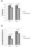
The U937 cell line was differentiated with phorbol esters and in vitro infected with L. infantum CM and HIFBS-depleted promastigotes at a phagocyte:promastigote ratio 1:20. The null hypothesis contrasted with the paired t-test was equal infectivity of HIFBS-depleted and CM promastigotes measured in triplicate as percentage of infected cells or number of amastigotes per infected cell. It was contrasted by the paired Student’s t-test (p-values provided in the graph). (A) Infection rate measured in terms of percentage of infected cells 24 and 48 h post-infection. Mean ± SD: 54 ± 1 (CM, 24 h); 54 ± 4 (CM, 48 h); 46 ± 3 (HIFBS-depletion, 48 h); 43 ± 1 (HIFBS-depletion, 48 h). (B) Average number of amastigotes per infected cell measured 24 and 48 h post-infection. Mean ± SD: 3.3 ± 0.3 (CM, 24 h); 5.0 ± 0.0 (CM, 48 h); 2.1 ± 0.1 (HIFBS-depletion, 48 h); 4.2 ± 0.1 (HIFBS-depletion, 48 h).
Fig 3
Fig 3. Genome microarray hybridization analysis for comparative expression profiling of HIFBS-depleted versus CM L. infantum promastigotes.
(A) Electropherograms (FU, fluorescence units vs. time in seconds) of HIFBS-depleted and CM promastigote total RNA triplicate samples obtained by capillary electrophoresis with the Agilent 2100 Bioanalyzer. (B) 1% agarose gel electrophoresis of amplified mRNA (aRNA) from total RNA samples of L. infantum HIFBS-depleted and CM promastigotes. (C) Average M/A scatter plots of the HIFBS-depletion/CM three-replicate microarray hybridization analysis. M = (log2Ri_log2Gi) and A = [(log2Ri + log2Gi)/2], where R and G are, respectively, red (Cy5-cDNA from HIFBS-depleted promastigotes) and green (Cy3-cDNA from CM) intensity values. Red spots correspond to selected DNA fragments containing a gene up-regulated at least 1.7 times and green spots represent those down-regulated at least 1.7 times. Further criteria for spot selection are detailed in the text. The SD values are displayed in the scatter plot. Differential expression was contrasted by the Student's t-test for each individual clone with the AlmaZen software. The statistically significant differences are highlighted in red and green as mentioned above.
Fig 4
Fig 4. LACK expression is maintained under serum depletion.
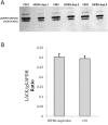
(A) Evaluation of relative abundance of the LACK antigen by Western blot performed with the anti-LACK polyclonal antibody (diluted 1:500) over total protein extracts of L. infantum promastigotes grown under HIFBS-depletion or in CM. The anti-gGAPDH polyclonal antibody (diluted 1:10,000) was used as the loading control antibody. The secondary antibody was HRP-conjugated goat anti-rabbit IgG (diluted 1:2,000). Chemoluminescence detection was performed with the ECL reagents (GE Healthcare) and the membrane was developed in an X-ray film. Upper bands are likely aggregates of the LACK protein [19]. (B) Bar graph of the relative expression ratio of the LACK antigen with respect to the gGAPDH. Mean and SD of three biological replicates are represented. Densitometry was performed with Gel Doc XR System and Quantity One version 4.6. software (BioRad). Both groups were compared by the Student's t-test including three biological replicates.
Fig 5
Fig 5. Differential gene expression triggered by serum depletion in culture.
Color legend: red, up-regulation in HIFBS-depleted promastigotes/CM; green, down-regulation. Genes involved in DNA repair, gene expression, protein folding, proteolysis, detoxification, signalling, the flagellum and the surface coat are up-regulated under serum depletion. The gene LinJ.31.1680 encodes the GINS complex protein subunit Psf3, which is involved in the initiation of DNA replication. GINS is a component of the eukaryotic replicative helicase essential for the establishment of DNA replication forks (reviewed in [51]). The transcript levels of this gene are more reduced in HIFBS-depleted promastigotes than in CM promastigotes. The hypothetical protein LinJ.24.1230 (hTFLi24.1230; sequence-specific DNA binding transcription factor activity GO0003700), the translation factor TFSUI1 and the DEAD-box helicase-like protein are up-regulated in HIFBS-depleted promastigotes, whereas the helicase is down-regulated. Two proteins involved in protein folding are also up-regulated (a cyclophilin and the heat shock protein 20, hsp20). Four genes involved in proteolysis are over-expressed: a calpain-like cysteine peptidase (C2Cp), a serine peptidase E, family 51 (SerP51), the proteasome non-ATPase regulatory subunit 8 (PSMD8) and the proteasome β subunit 3 (PSMB3). PSMB3 and PSMD8 are involved in protein degradation via the ubiquitin-proteasome system. The surface molecule-encoding genes hydrophilic surface protein B (HASPB) and amastin-like LinJ.34.2660 are up-regulated, as well as the glyoxylase I (GLO1), one of the components of the glyoxylase detoxification system.
Fig 6
Fig 6. Enzyme activity of branched chain amino transferase in total protein extracts.
(A) Basis of the BCAT assay. Enzymes and reagents added to the protein extracts are represented in bold. Ile transamination is reversible and the α-ketoglutarate is the amino acceptor. In this assay, the reverse reaction takes place because the 3-methyl-2-oxopentanoic acid is provided as the amino acceptor and Glu as the donor. This reaction is coupled to Asp transamination by adding this substrate and the ASAT. The product of this reaction (OAA) is reduced to malate by the MDH in the presence of the coenzyme NADH. The BCAT activity is indirectly measured as NADH oxidation (absorbance decay at 334 nm). Abbreviations: ASAT, L-aspartate aminotransferase; Glu, glutamic acid; MDH, malate dehydrogenase; PLP, pyridoxal phosphate. (B) BCAT activity in protein extracts of HIFBS-depleted and CM promastigotes. Three biological replicates were performed. HIFBS-depletion significantly decreases the specific BCAT enzymatic activity (EA) (t-test, p* = 0.022 < 0.05). Means and standard deviation (SD) of the EA are provided.

Similar articles

Cited by

References

    1. Arce A, Estirado A, Ordobas M, Sevilla S, Garcia N, Moratilla L, et al. Re-emergence of leishmaniasis in Spain: community outbreak in Madrid, Spain, 2009 to 2012. Euro Surveill. 2013;18(30):20546 Epub 2013/08/10. 20546 [pii]. . - PubMed
    1. Jimenez M, Gonzalez E, Martin-Martin I, Hernandez S, Molina R. Could wild rabbits (Oryctolagus cuniculus) be reservoirs for Leishmania infantum in the focus of Madrid, Spain? Vet Parasitol. 2014;202(3–4):296–300. Epub 2014/04/30. 10.1016/j.vetpar.2014.03.027 S0304-4017(14)00202-7 [pii]. . - DOI - PubMed
    1. Molina R, Jimenez MI, Cruz I, Iriso A, Martin-Martin I, Sevillano O, et al. The hare (Lepus granatensis) as potential sylvatic reservoir of Leishmania infantum in Spain. Vet Parasitol. 2012;190(1–2):268–71. Epub 2012/06/09. 10.1016/j.vetpar.2012.05.006 S0304-4017(12)00258-0 [pii]. . - DOI - PubMed
    1. Killick-Kendrick R. The biology and control of phlebotomine sand flies. Clin Dermatol. 1999;17(3):279–89. Epub 1999/06/29. S0738-081X(99)00046-2 [pii]. . - PubMed
    1. Bates PA. Housekeeping by Leishmania. Trends Parasitol. 2006;22(10):447–8. Epub 2006/08/15. S1471-4922(06)00204-2 [pii] 10.1016/j.pt.2006.08.003 - DOI - PMC - PubMed

Publication types

MeSH terms