

Intraductal papillary mucinous neoplasms of the pancreas: radiological predictors of malignant transformation and the introduction of bile duct dilation to current guidelines
- PMID: 26959611
- PMCID: PMC4985463
- DOI: 10.1259/bjr.20150853
Intraductal papillary mucinous neoplasms of the pancreas: radiological predictors of malignant transformation and the introduction of bile duct dilation to current guidelines
Abstract
Objective: To evaluate the current guidelines as a model to predict malignancy and to determine further radiological predictors of malignancy in intraductal papillary mucinous neoplasms (IPMNs).
Methods: 384 patients who had undergone a pancreatic operation with the pathological diagnosis of IPMN as well as applicable pre-operative imaging (CT/MRI) were included in the study. Images were evaluated retrospectively in consensus by two radiologists, using a standardized checklist. Descriptive statistics, binary logistic regression and receiver operator curve analysis were performed to assess the International Consensus Guidelines and other radiological predictors of clinical malignancy (defined as carcinoma in situ and invasive carcinoma).
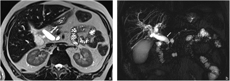
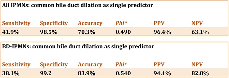
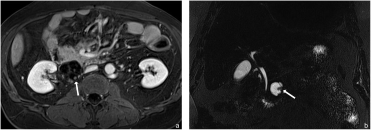
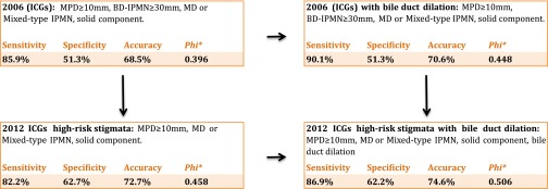
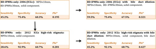
Results: The best independent predictors of malignancy (n = 191) were solid components [odds ratio (OR) 3.98], parenchymal atrophy with main pancreatic duct dilation 5-9 mm (OR: 5.1) and common bile duct (CBD) dilation (OR: 31.26). >96% of all cases with CBD dilation were malignant IPMNs (positive-predictive value 96.4%; negative-predictive value 63.1%). Analysis of the current guidelines showed a diagnostic improvement with the addition of CBD dilation on determining the malignancy of IPMNs (sensitivity 82.2%/86.9%; specificity 72.7%/74.6%). Subanalysis of branch duct intraductal papillary mucinous neoplasms (BD-IPMNs; n = 168) also resulted in a diagnostic improvement with the addition of CBD dilation (sensitivity 28.6%/45.2%; specificity 92.9%/92.1%). The best independent predictors of malignancy for BD-IPMNs were parenchymal atrophy (OR: 4.00) and CBD dilation (OR: 29.3). Frequency analysis revealed that even small BD-IPMNs had already undergone malignant transformation (≤1 cm: 15%; 1-2 cm: 26%; 2-3 cm: 20%) with about 10% of those having a dilated bile duct.
Conclusion: CBD dilation was a significant positive predictor of malignancy in IPMNs regardless of their size.
Advances in knowledge: Introduction of CBD dilation as a radiological predictor for malignancy might increase the diagnostic accuracy of current imaging-based guidelines.
Figures
References
-
- Klöppel G, Solcia E, Longnecker D, Capella C, Sobin L. Histological typing of tumors of the exocrine pancreas. 2nd edn. Berlin, Germany: Springer; 1996.
MeSH terms
LinkOut - more resources
Full Text Sources
Other Literature Sources
Medical
