Comment
. 2016 Jun;23(6):510-9.
doi: 10.1038/gt.2016.23. Epub 2016 Apr 7.

Ultrasound-targeted hepatic delivery of factor IX in hemophiliac mice

Affiliations
Comment

Ultrasound-targeted hepatic delivery of factor IX in hemophiliac mice

C D Anderson et al. Gene Ther. 2016 Jun.

Abstract

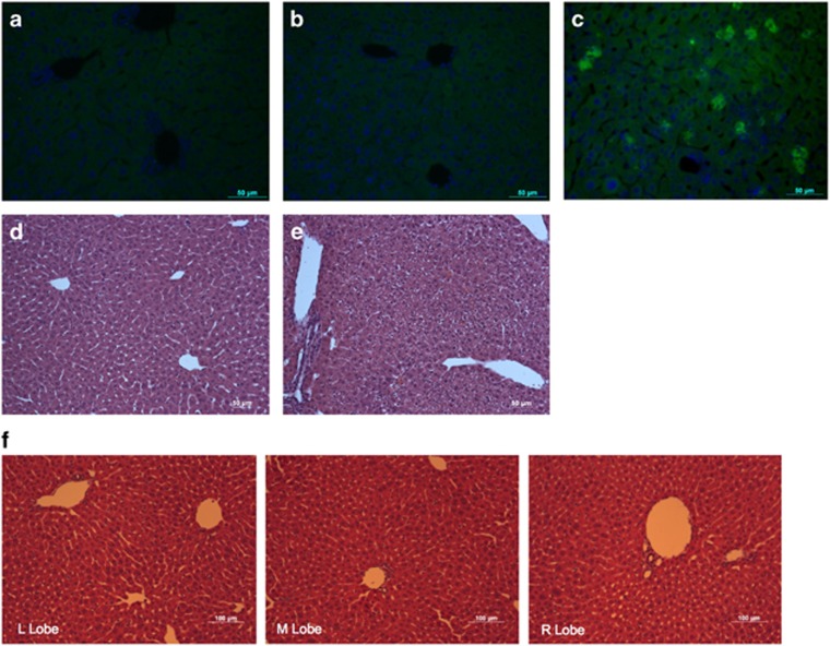
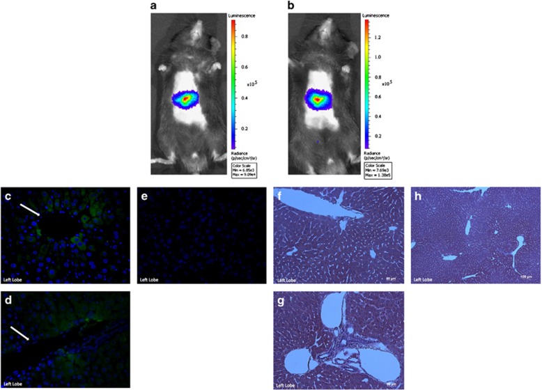
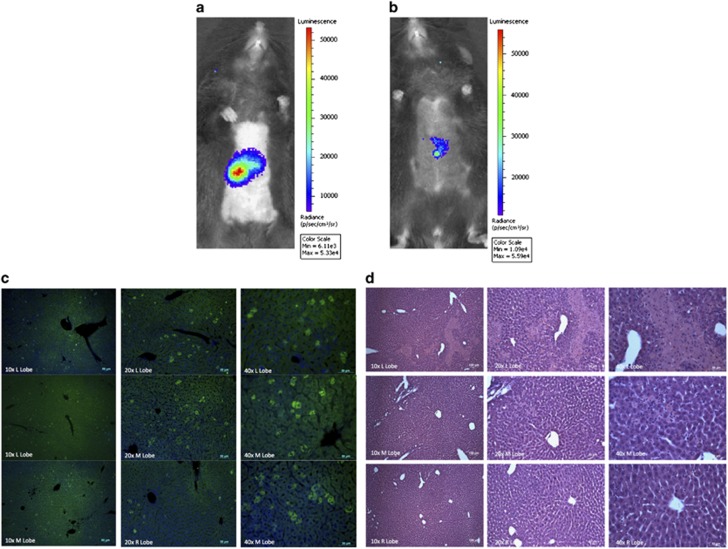
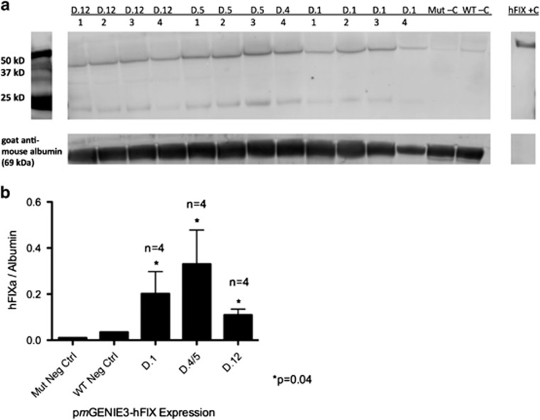
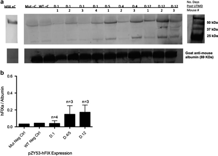
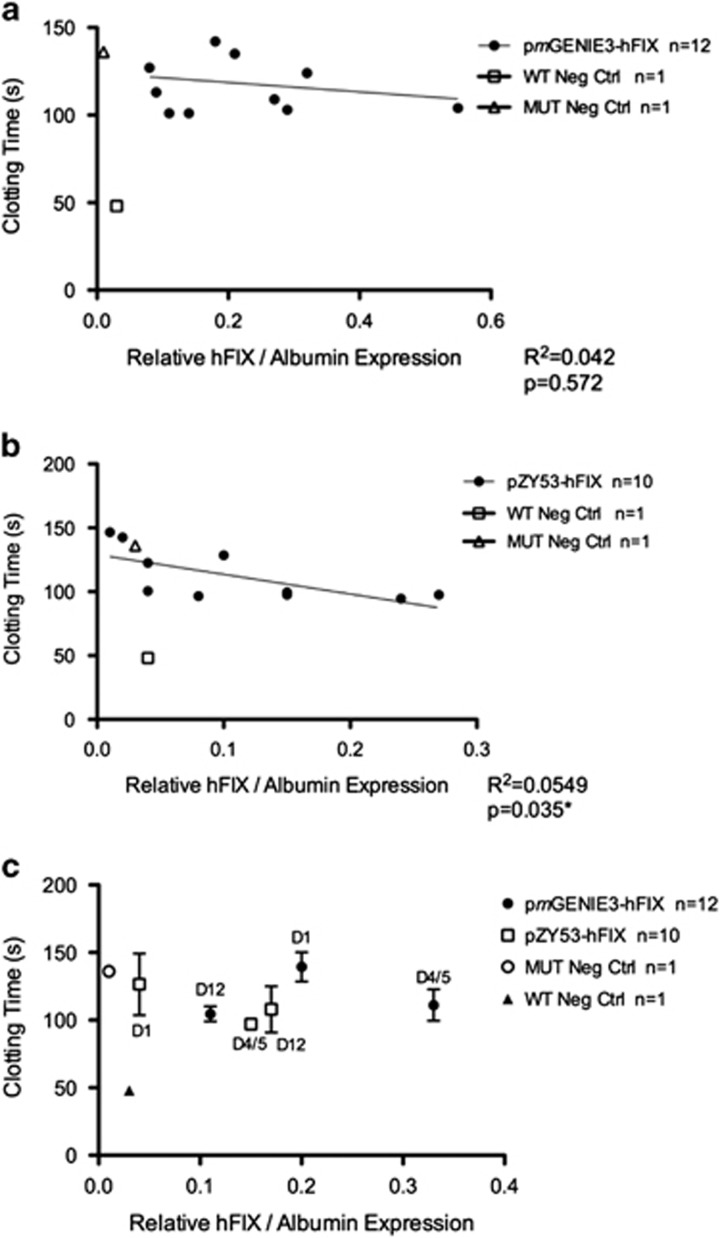
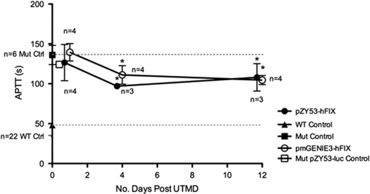
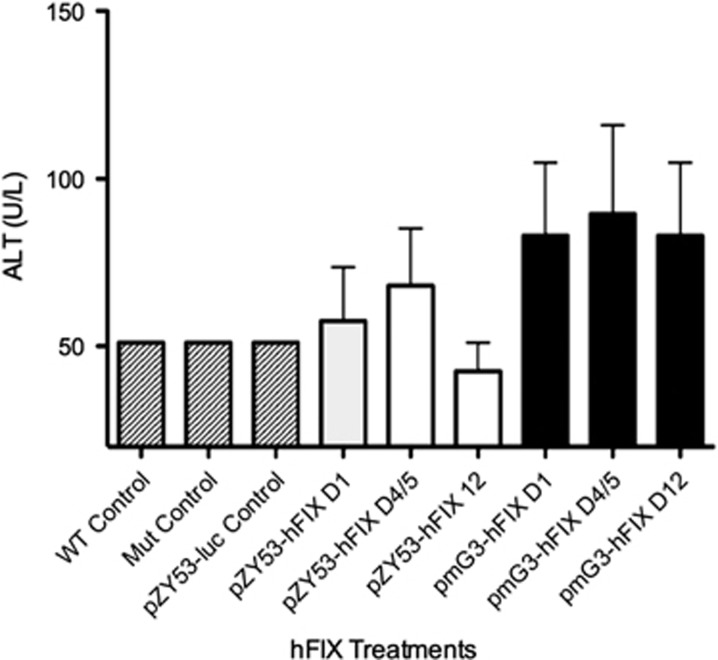
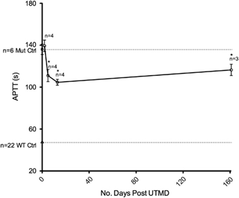
Ultrasound-targeted microbubble destruction (UTMD) was used to direct the delivery of plasmid and transposase-based vectors encoding human factor IX (hFIX) to the livers of hemophilia B (FIX-/-) mice. The DNA vectors were incorporated into cationic lipid microbubbles, injected intravenously, and transfected into hepatocytes by acoustic cavitation of the bubbles as they transited the liver. Ultrasound parameters were identified that produced transfection of hepatocytes in vivo without substantial damage or bleeding in the livers of the FIX-deficient mice. These mice were treated with a conventional expression plasmid, or one containing a piggyBac transposon construct, and hFIX levels in the plasma and liver were evaluated at multiple time points after UTMD. We detected hFIX in the plasma by western blotting from mice treated with either plasmid during the 12 days after UTMD, and in the hepatocytes of treated livers by immunofluorescence. Reductions in clotting time and improvements in the percentage of FIX activity were observed for both plasmids, conventional (4.15±1.98%), and transposon based (2.70±.75%), 4 to 5 days after UTMD compared with untreated FIX (-/-) control mice (0.92±0.78%) (P=0.001 and P=0.012, respectively). Reduced clotting times persisted for both plasmids 12 days after treatment (reflecting percentage FIX activity of 3.12±1.56%, P=0.02 and 3.08±0.10%, P=0.001, respectively). Clotting times from an additional set of mice treated with pmGENIE3-hFIX were evaluated for long-term effects and demonstrated a persistent reduction in average clotting time 160 days after a single treatment. These data suggest that UTMD could be a minimally invasive, nonviral approach to enhance hepatic FIX expression in patients with hemophilia.


Conflict of interest statement

Stefan Moisyadi is the owner of Manoa BioSciences, a start-up company out of the University of Hawaii. Manoa BioSciences holds the rights and patent for the pmGENIE construct.

Figures

Figure 1
Figure 1
Histological discrimination of human FIX expression from mouse FIX and liver histology of wild-type and mutant control mice. (a) Untreated wild-type mouse left liver lobe stained with hFIX antibody, × 40. (b) Untreated FIX mutant mouse left liver lobe stained with hFIX antibody, × 40. (c) Human FIX UTMD-treated mutant mouse left liver lobe stained with hFIX antibody at 1 day after treatment, × 40. (d, e) Immunofluorescence and H&E-stained left liver lobes from untreated mice demonstrate the absence of hFIX expression and antibody crossreactivity to murine FIX (mFIX). (f) hFIX negative control histology. H&E stain of a mutant mouse liver 1 day after treatment with pZY53-luc and microbubbles (without hFIX) followed by UTMD.
Figure 2
Figure 2
Co-delivery of liver-specific hFIX and reporter vectors to the mutant mouse liver. (a, b) Expression from pZY53-luc (co-delivered with pZY53-hFIX) 2 (a) and 5 (b) days after UTMD. Luciferase expression (luc) is measured as the average radiance in units of photons per s per cm2 per steradian. (c–e) hFIX expression in the mutant mouse liver by immunofluorescence. Left liver lobe immunofluorescence from treated mutant mouse 5 days after UTMD-mediated delivery of pZY53-hFIX. Hepatocytes expressing the hFIX transgene appear green and were incubated with a goat anti-human FIX primary antibody and then stained with an Alexa Fluor 488 fluorescent-conjugated secondary antibody, and nuclei are stained blue with DAPI, × 40 magnification (c, d). White arrows indicate hFIX transgene expression surrounding hepatic blood vessels. Negative control (e), liver section stained only with the secondary antibody and DAPI. (f–h) H&E staining of mutant mouse liver 5 days after UTMD-mediated delivery of hFIX (pZY53-hFIX). Left liver lobe sections, scale=50 μm (f, g) and 100 μm (h).
Figure 3
Figure 3
Co-delivery of liver-specific hFIX and reporter vectors to the mutant mouse liver. (a, b) Expression from pZY53-luc (co-delivered with pmGENIE3-hFIX) at 1 (a) and 12 (b) days after UTMD. Luciferase expression (luc) is measured as the average radiance in units of photons per s per cm2 per steradian. (c) Immunofluorescence images of hFIX expression in the left and medial lobes (× 10–40) of the mutant mouse liver 1 day after treatment with pmGENIE3-hFIX. (d) H&E staining of mutant mouse liver 1 day after UTMD-mediated delivery of hFIX (pmGENIE3-hFIX). Top panel, left liver lobe sections (× 10–40), middle panel, medial liver lobe sections (× 10–40), and bottom panel, right liver lobe sections (× 10–40), scale=100 μm (× 10) and 50 μm (× 20–40).
Figure 4
Figure 4
hFIX expression in mutant mouse plasma. Mice treated with pmGENIE3-hFIX and samples collected 1 (n=4), 4 or 5 (n=4) and 12 (n=4) days after UTMD. (a) Blot shown with hFIX recombinant protein positive control. Mut–C, untreated mutant plasma negative control; WT–C, untreated wild-type plasma negative control. Goat anti-mouse serum albumin antibody was used as a loading control. (b) Densitometric analysis of pmGENIE3-hFIX expression relative to mouse anti-serum albumin loading control.
Figure 5
Figure 5
hFIX expression in mutant mouse plasma. Mice treated with pZY53-hFIX and samples collected 1 (n=4), 4 or 5 (n=3) and 12 (n=3) days after UTMD. (a) Blot shown with hFIX recombinant protein positive control. Mut–C, untreated mutant plasma negative control; WT –C, untreated wild-type plasma negative control. Goat anti-mouse serum albumin antibody was used as a loading control. (b) Densitometric analysis of pZY53-hFIX expression relative to mouse anti-serum albumin loading control.
Figure 6
Figure 6
Relative hFIX expression compared with clotting times. (a) Relative hFIX expression versus clotting time for mice treated with pmGENIE3-hFIX. (b) Relative hFIX expression versus clotting time for mice treated with pZY53-hFIX. (c) Average hFIX expression and clotting times for each treatment time point for pmGENIE3-hFIX and pZY53-hFIX.
Figure 7
Figure 7
APTT values for UTMD hFIX-treated mutant mice. Average clotting times for three time points tested for each group of mutant mice treated with pZY53-hFIX or pmGENIE3-hFIX by UTMD (n=3–4 mice per group, as indicated). Dashed lines indicate baseline average clotting times for untreated wild-type and mutant mice. One mutant control mouse (indicated by open square) was treated with pZY53-luc plasmid only by UTMD and APTT test was performed on plasma 1 day after UTMD as a negative control for hFIX. P=0.001 and P=0.02 for pZY53-hFIX, P=0.012 and P=0.001 for pmGENIE3-hFIX days 4–5 and 12 after UTMD, respectively.
Figure 8
Figure 8
ALT assay on treated mutant mouse plasma. Average ALT levels 1, 4 or 5 and 12 days after UTMD treatment with pZY53-hFIX (n=4 for day 1 and n=3 for days 4 or 5 and 12 time points) or pmGENIE3-hFIX (n=4 per time point). Untreated wild-type, mutant and pZY53-luc-treated mouse plasma served as controls (n=1 per control group).
Figure 9
Figure 9
Long-term APTT evaluation in mutant mice treated with pmGENIE3-hFIX. Open circles indicate average clotting times in mice 1, 4 or 5, 12 and 160 days after treatment with pmGENIE3-hFIX (n=3–4 mice per group, as indicated). Dashed lines indicate baseline average clotting times for untreated wild-type (n=22, black triangle) and mutant (n=6, black square) mice. P=0.012, P=0.001 and P=0.044, days 4 or 5, 12 and 160 after UTMD, respectively, when compared with untreated mutant controls.

Comment on

  • Adenovirus-associated virus vector-mediated gene transfer in hemophilia B.
    Nathwani AC, Tuddenham EG, Rangarajan S, Rosales C, McIntosh J, Linch DC, Chowdary P, Riddell A, Pie AJ, Harrington C, O'Beirne J, Smith K, Pasi J, Glader B, Rustagi P, Ng CY, Kay MA, Zhou J, Spence Y, Morton CL, Allay J, Coleman J, Sleep S, Cunningham JM, Srivastava D, Basner-Tschakarjan E, Mingozzi F, High KA, Gray JT, Reiss UM, Nienhuis AW, Davidoff AM. Nathwani AC, et al. N Engl J Med. 2011 Dec 22;365(25):2357-65. doi: 10.1056/NEJMoa1108046. Epub 2011 Dec 10. N Engl J Med. 2011. PMID: 22149959 Free PMC article. Clinical Trial.
  • Long-term safety and efficacy of factor IX gene therapy in hemophilia B.
    Nathwani AC, Reiss UM, Tuddenham EG, Rosales C, Chowdary P, McIntosh J, Della Peruta M, Lheriteau E, Patel N, Raj D, Riddell A, Pie J, Rangarajan S, Bevan D, Recht M, Shen YM, Halka KG, Basner-Tschakarjan E, Mingozzi F, High KA, Allay J, Kay MA, Ng CY, Zhou J, Cancio M, Morton CL, Gray JT, Srivastava D, Nienhuis AW, Davidoff AM. Nathwani AC, et al. N Engl J Med. 2014 Nov 20;371(21):1994-2004. doi: 10.1056/NEJMoa1407309. N Engl J Med. 2014. PMID: 25409372 Free PMC article. Clinical Trial.

References

    1. Snyder RO, Miao C, Meuse L, Tubb J, Donahue BA, Lin HF et al. Correction of hemophilia B in canine and murine models using recombinant adeno-associated viral vectors. Nat Med 1999; 5: 64–70. - PubMed
    1. Snyder RO, Miao CH, Patijn GA, Spratt SK, Danos O, Nagy D et al. Persistent and therapeutic concentrations of human factor IX in mice after hepatic gene transfer of recombinant AAV vectors. Nat Genet 1997; 16: 270–276. - PubMed
    1. Monahan PE. Factor IX: insights from knock-out and genetically engineered mice. Thromb Haemost 2008; 100: 563–575. - PubMed
    1. Nathwani AC, Tuddenham EG, Rangarajan S, Rosales C, McIntosh J, Linch DC et al. Adenovirus-associated virus vector-mediated gene transfer in hemophilia B. N Engl J Med 2011; 365: 2357–2365. - PMC - PubMed
    1. Miao CH. Advances in overcoming immune responses following hemophilia gene therapy. J Genet Syndr Gene Ther 2011; S1: 007. - PMC - PubMed

Publication types