. 2016 Apr 7;18(13):8911-8919.
doi: 10.1039/c6cp00476h.

Combined probes of X-ray scattering and optical spectroscopy reveal how global conformational change is temporally and spatially linked to local structural perturbation in photoactive yellow protein

Affiliations

Combined probes of X-ray scattering and optical spectroscopy reveal how global conformational change is temporally and spatially linked to local structural perturbation in photoactive yellow protein

Tae Wu Kim et al. Phys Chem Chem Phys. .

Abstract

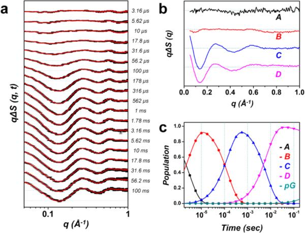
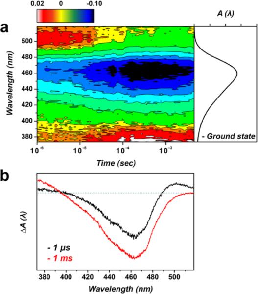
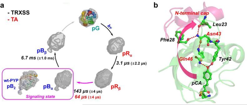
Real-time probing of structural transitions of a photoactive protein is challenging owing to the lack of a universal time-resolved technique that can probe the changes in both global conformation and light-absorbing chromophores of the protein. In this work, we combine time-resolved X-ray solution scattering (TRXSS) and transient absorption (TA) spectroscopy to investigate how the global conformational changes involved in the photoinduced signal transduction of photoactive yellow protein (PYP) is temporally and spatially related to the local structural change around the light-absorbing chromophore. In particular, we examine the role of internal proton transfer in developing a signaling state of PYP by employing its E46Q mutant (E46Q-PYP), where the internal proton transfer is inhibited by the replacement of a proton donor. The comparison of TRXSS and TA spectroscopy data directly reveals that the global conformational change of the protein, which is probed by TRXSS, is temporally delayed by tens of microseconds from the local structural change of the chromophore, which is probed by TA spectroscopy. The molecular shape of the signaling state reconstructed from the TRXSS curves directly visualizes the three-dimensional conformations of protein intermediates and reveals that the smaller structural change in E46Q-PYP than in wild-type PYP suggested by previous studies is manifested in terms of much smaller protrusion, confirming that the signaling state of E46Q-PYP is only partially developed compared with that of wild-type PYP. This finding provides direct evidence of how the environmental change in the vicinity of the chromophore alters the conformational change of the entire protein matrix.


Figures

Fig. 1
Fig. 1
Structure of E46Q-PYP. Structures of the protein backbone (ribbon) and the active site around the chromophore (ball-and-stick) in E46Q-PYP (PDB: 1OTA). In E46Q-PYP, the glutamic acid (Glu46) in wild-type PYP was replaced by a glutamine (Gln46). The dashed lines indicate the hydrogen bonds between p-coumaric acid (pCA) and the neighbouring residues in the ground state of E46Q. Due to the effect of mutation, the hydrogen bond between pCA and Gln46 is longer than the one between pCA and Glu46 in wild-type PYP.
Fig. 2
Fig. 2
Time-resolved X-ray solution scattering data of the E46Q-PYP photocycle and global kinetic analysis. (a) Experimental (black) and theoretical (red) difference scattering curves. (b) Species-associated difference scattering curves extracted from the PCA analysis. (c) Time-dependent population change of the intermediates following the proposed kinetic model determined by the PCA analysis.
Fig. 3
Fig. 3
Transient absorption spectra of E46Q-PYP. (a) The contour shows the time evolution of transient absorption spectra. The ground state absorption spectrum of E46Q-PYP is shown together on the right side of the TA spectra. At ~64 μs, the decay of the positive feature at ~500 nm concurs with the growth of positive signal on ~380 nm, which is the signature of the spectrally blue-shifted intermediate called pB. (b) TA spectra at two representative time delays, 1 μs and 1 ms.
Fig. 4
Fig. 4
Structural dynamics of E46Q-PYP photocycle and hydrogen-bonding network from the chromophore binding pocket to the N-terminal cap. (a) Global kinetics of E46Q-PYP photocycle. The reconstructed molecular shapes of the intermediates were extracted from the structural analysis of the scattering data. The photocycle of E46Q-PYP includes four intermediates (pRα, pRβ, pBα, and pBβ) and the transition rates among the intermediates were determined from time-resolved X-ray solution scattering (TRXSS) and transient absorption spectroscopy (TA). The global conformational change of the protein is maximal in the pRβ → pBα transition occurring on the time scale of 143 μs as supported by the reconstructed shape from TRXSS. The inset (magenta box) shows the reconstructed molecular shape of the signaling state (pB2) of wt-PYP with the atomistic structure determined from the previous study combining DEER, NMR, and SAXS/WAXS experiment. (b) The local structural change in the vicinity of chromophore (pCA) can be propagated up to the N-terminal cap region through the hydrogen-bonding network (red dashed lines). The weak hydrogen bond between pCA and Gln46 in E46Q PYP may result in the smaller global conformational change compared with the change in wild-type PYP.

References

    1. Kim JG, Kim TW, Kim J, Ihee H. Acc. Chem. Res. 2015;48:2200–2208. - PMC - PubMed
    1. Bredenbeck J, Helbing J, Kumita JR, Woolley GA, Hamm P. Proc. Natl. Acad. Sci. U.S.A. 2005;102:2379–2384. - PMC - PubMed
    1. Schotte F, Lim MH, Jackson TA, Smirnov AV, Soman J, Olson JS, Phillips GN, Wulff M, Anfinrud PA. Science. 2003;300:1944–1947. - PubMed
    1. Chen EF, Kumita JR, Woolley GA, Kliger DS. J. Am. Chem. Soc. 2003;125:12443–12449. - PubMed
    1. Lewis JW, Goldbeck RA, Kliger DS, Xie XL, Dunn RC, Simon JD. J. Phys. Chem. 1992;96:5243–5254.

Publication types

MeSH terms

Substances

LinkOut - more resources