Review
. 2016 Apr;29(2):401-27.
doi: 10.1128/CMR.00069-15.

Clinical Significance and Pathogenesis of Staphylococcal Small Colony Variants in Persistent Infections

Affiliations
Review

Clinical Significance and Pathogenesis of Staphylococcal Small Colony Variants in Persistent Infections

Barbara C Kahl et al. Clin Microbiol Rev. 2016 Apr.

Abstract

Small colony variants (SCVs) were first described more than 100 years ago for Staphylococcus aureus and various coagulase-negative staphylococci. Two decades ago, an association between chronic staphylococcal infections and the presence of SCVs was observed. Since then, many clinical studies and observations have been published which tie recurrent, persistent staphylococcal infections, including device-associated infections, bone and tissue infections, and airway infections of cystic fibrosis patients, to this special phenotype. By their intracellular lifestyle, SCVs exhibit so-called phenotypic (or functional) resistance beyond the classical resistance mechanisms, and they can often be retrieved from therapy-refractory courses of infection. In this review, the various clinical infections where SCVs can be expected and isolated, diagnostic procedures for optimized species confirmation, and the pathogenesis of SCVs, including defined underlying molecular mechanisms and the phenotype switch phenomenon, are presented. Moreover, relevant animal models and suggested treatment regimens, as well as the requirements for future research areas, are highlighted.


Figures

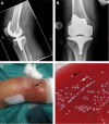
FIG 1
FIG 1
Periprosthetic knee joint infection of a patient with a history of clinical and infectious laboratory parameters for 2 weeks. A two-stage revision was performed. (A and B) X-rays without specific changes. (C) Clinical presentation 2 to 3 weeks after the first symptoms occurred. Inflamed skin with draining pus after incision of the skin (black arrow) can be seen. (D) S. aureus colonies grown from a specimen of chronically infected bone tissue. On the Columbia blood agar plate, a high phenotypic diversity of large (black arrow), small, and very small (white arrow) phenotypes, which represent dynamic SCVs, is visible. (Panels A, B, and C courtesy of W. Kluge, Hufeland-Klinikum Weimar, Germany; reprinted with permission.)
FIG 2
FIG 2
S. aureus recovered from the airways of a cystic fibrosis patient. (A) Gram staining of a sputum specimen from a CF patient. Gram-positive cocci (black arrow) representative of S. aureus and numerous neutrophils (white arrow) are visible. (B) Mixture of large (black arrow) and small (white arrow) colonies cultured from a sputum sample, indicative of a mixture of normal S. aureus and S. aureus SCVs, on Columbia blood agar. (C) Growth of large (black arrow) and small (white arrow) colonies typical for a mixture of normal S. aureus and S. aureus SCVs on SAID chromogenic agar, used as a selective agar for S. aureus, from a sputum sample from a CF patient. (D) Pulsed-field gel electrophoresis of normal S. aureus and S. aureus SCVs, which persisted in the airways of an individual CF patient from 1994 until 2000. n, normal S. aureus; S, SCV; lane 1, marker; lane 2, normal S. aureus (NS) 6/1994; lanes 3 and 4, SCV and NS isolated together in September 1995; lanes 5 and 6, SCV and NS isolated together in June 1995; lanes 7 and 8, SCV and NS isolated in January 1997; lane 9, SCV from May 1998; lane 10, SCV from October 1999; lane 11, SCV from January 2000; lane 12, marker.
FIG 3
FIG 3
Scheme of dynamic SCV development in the course from acute to chronic infection. During acute infection, bacteria show high-level expression of agr and regulated toxins causing inflammatory and cytotoxic reactions. In the chronic course of the infection, bacteria apply adaptation mechanisms to resist cellular degradation that involve formation of dynamic SCV-like phenotypes, downregulation of agr, and upregulation of SigB. When leaving the intracellular shelter, the SCVs can rapidly revert back to the fully aggressive wild-type phenotype, which can cause a new episode of infection.
FIG 4
FIG 4
Overview of the menaquinone heme and thymidine pathways. Enzymes encoded by genes known to lead to the SCV phenotype if disabled are shown in red (knockout-generated SCVs are indicated with rectangular boxes, and naturally occurring or gentamicin-induced SCVs are indicated with ovals). DHN, 1,4-dihydroxy-2-naphthoate; GLDH, glutamate dehydrogenase; GltX, glutamate-tRNA ligase; HemA, glutamyl-tRNA reductase; HemB, 5-aminolevulinic acid dehydratase (porphobilinogen synthase); HemC, porphobilinogen deaminase; HemD, uroporphyrinogen III synthase; HemE, uroporphyrinogen decarboxylase; HemF, coproporphyrinogen III oxidase; HemG, protoporphyrinogen oxidase; HemH, protoheme ferrolyase (ferrochelatase); HemL, glutamate-1-semialdehyde aminotransferase; HemN, oxygen-independent coproporphyrinogen III oxidase; HemY, oxygen-dependent protoporphyrinogen oxidase; HSDC, (1R,6R)-6-hydroxy-2-succinylcyclohexa-2,4-diene-1-carboxylate; ICDH, isocitrate dehydrogenase; MenA, 1,4-dihydroxy-2-naphthoate octaprenyltransferase; MenB, 1,4-dihydroxy-2-naphthoyl-CoA synthase; MenC, o-succinylbenzoate synthase; MenD, SEPHCHC synthase; MenE, 2-succinylbenzoate-CoA ligase; MenF, isochorismate synthase; MenG, demethylmenaquinone methyltransferase; MenH, SHCHC synthase (SHCHC synthase activity was previously attributed to MenD); SEPHCHC, 2-succinyl-5-enolpyruvyl-6-hydroxy-3-cyclohexene-1-carboxylate; SHCHC, (1R,6R)-2-succinyl-6-hydroxy-2,4-cyclohexadiene-1-carboxylate; TYMK, deoxythymidylate kinase; ThyA (TYMS), thymidylate synthase. Pathways are according to the Kyoto Encyclopedia of Genes and Genomes (KEGG), release 74.0 (http://www.genome.jp/kegg/).
None
None
None

References

    1. Kolle W, Hetsch H. 1906. Die experimentelle Bakteriologie und die Infektionskrankheiten mit besonderer Berücksichtigung der Immunitätslehre. Ein Lehrbuch für Studierende, Ärzte und Medizinalbeamte. Urban & Schwarzenberg, Berlin, Germany.
    1. Hoffstadt RE, Youmans GP. 1934. The genetic significance of the dissociants of Staphylococcus aureus. J Bacteriol 27:551–560. - PMC - PubMed
    1. Borderon E, Horodniceanu T. 1976. Mutants déficients a colonies naines de Staphylococcus: étude de trois souches isolées chez des malades porteurs d'ostéosynthèses. Ann Microbiol (Paris) 127:503–514. - PubMed
    1. Goudie JB, Goudie RB. 1955. Recurrent infection by a stable-dwarf-colony variant of Staphylococcus aureus. J Clin Pathol 8:284–287. doi:10.1136/jcp.8.4.284. - DOI - PMC - PubMed
    1. Spink WW, Osterberg K, Finstad J. 1962. Human endocarditis due to a strain of CO2-dependent penicillin-resistant staphylococcus producing dwarf colonies. J Lab Clin Med 59:613–619. - PubMed

Publication types

MeSH terms

LinkOut - more resources