Review
. 2016 Jun;74(4):ftw018.
doi: 10.1093/femspd/ftw018.

Pathogenesis of Candida albicans biofilm

Review

Pathogenesis of Candida albicans biofilm

Christina Tsui et al. Pathog Dis. 2016 Jun.

Abstract

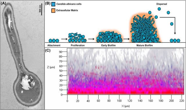
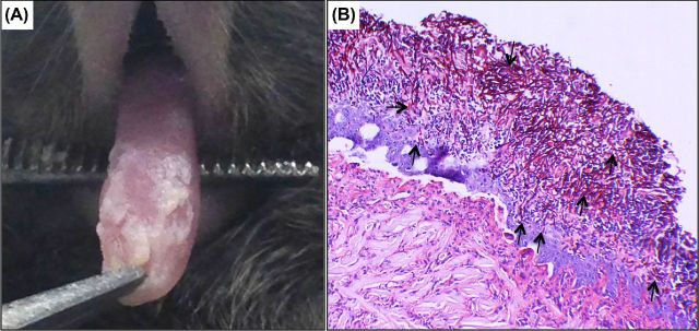
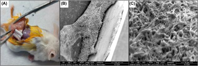
Candida albicans is the most common human fungal pathogen causing diseases ranging from mucosal to systemic infections. As a commensal, C. albicans asymptomatically colonizes mucosal surfaces; however, any disruption in the host environment or under conditions of immune dysfunction, C. albicans can proliferate and invade virtually any site in the host. The ability of this highly adaptable fungal species to transition from commensal to pathogen is due to a repertoire of virulence factors. Specifically, the ability to switch morphology and form biofilms are properties central to C. albicans pathogenesis. In fact, the majority of C. albicans infections are associated with biofilm formation on host or abiotic surfaces such as indwelling medical devices, which carry high morbidity and mortality. Significantly, biofilms formed by C. albicans are inherently tolerant to antimicrobial therapy and therefore, the susceptibility of Candida biofilms to the current therapeutic agents remains low. The aim of this review is to provide an overview of C. albicans highlighting some of the diverse biofilm-associated diseases caused by this opportunistic pathogen and the animal models available to study them. Further, the classes of antifungal agents used to combat these resilient infections are discussed along with mechanisms of drug resistance.


Figures

Figure 1.
Figure 1.
Candida albicans morphogenesis and biofilm development. (A) Transmission electron micrograph of a germinating yeast cell. Germ tube can be seen as a filament, which continues to elongate forming a hypha. (B) A schematic illustrating the stages of C. albicans biofilm development. Adherence: yeast cells adhere to a substrate forming a yeast basal layer. Initiation: cells propagate and form germ tubes. Maturation: hyphae are formed and extracellular matrix accumulates. Dispersal: cells are released from the biofilm and are dispersed to seed new locations. (C) Confocal scanning laser microscopy of in vitro grown C. albicans biofilm; 48 h biofilm stained with FUN1 (blue) vital fungal stain and the biofilm polysaccharide matrix stain Concanavalin A (ConA) (red). Images were obtained using a Zeiss 710 confocal microscope by LSM 5 Image Browser software at a resolution of 512 × 512 pixels, with an average of 8 images per line. A series of images at ≤1 μm intervals in the z-axis were acquired for the full depth of the biofilm.
Figure 2.
Figure 2.
Oral candidiasis. Clinical manifestation of oral candidiasis in a human host characterized by white lesions formed on oral mucosal surfaces: (A) buccal mucosa (B) palate and (C) gingiva.
Figure 3.
Figure 3.
Mouse model of oral candidiasis. (A) Four days post sublingual infection with C. albicans, mice develop oral candidiasis characterized by white plaques covering the tongue and other oral surfaces. (B) Histopathology of tongue tissue from mouse with oral candidiasis demonstrating extensive penetration of epithelial tissue by the invasive C. albicans hyphae (arrows).
Figure 4.
Figure 4.
Mouse subcutaneous catheter model. (A) Explant of subcutaneous catheters from mouse 48 h post implantation. Candida albicans 48 h biofilm grown in vivo in the lumen of catheters. (B) Scanning electron micrographs demonstrating the thick hyphal matrix formed in the lumen of the recovered implanted catheters. (C) High magnification image of the formed biofilm showing the extracellular matrix material, which appears fibrous.

References

    1. Al-Fattani MA, Douglas LJ. Biofilm matrix of Candida albicans and Candida tropicalis: chemical composition and role in drug resistance. J Med Microbiol. 2006;55:999–1008. - PubMed
    1. Andes D, Nett J, Pschel P, et al. Development and characterization of an in vivo central venous catheter Candida albicans biofilm model. Infect Immun. 2004;72:6023–31. - PMC - PubMed
    1. Bachmann SP, VandeWalle K, Ramage G, et al. In vitro activity of caspofungin against Candida albicans biofilm. Antimicrob Agents Ch. 2002;46:3591–6. - PMC - PubMed
    1. Baillie GS, Douglas J. Effect of growth rate on resistance of Candidaalbicans biofilms to antifungal agents. Antimicrob Agents Ch. 1998a;42:1900–5. - PMC - PubMed
    1. Baillie GS, Douglas J. Iron-limited biofilms of Candidaalbicans and their susceptibility to amphotericin B. Antimicrob Agents Ch. 1998b;42:2146–9. - PMC - PubMed

Publication types

MeSH terms