. 2016 Mar 10:6:22850.
doi: 10.1038/srep22850.

Human malignant mesothelioma is recapitulated in immunocompetent BALB/c mice injected with murine AB cells

Affiliations

Human malignant mesothelioma is recapitulated in immunocompetent BALB/c mice injected with murine AB cells

Rosanna Mezzapelle et al. Sci Rep. .

Abstract

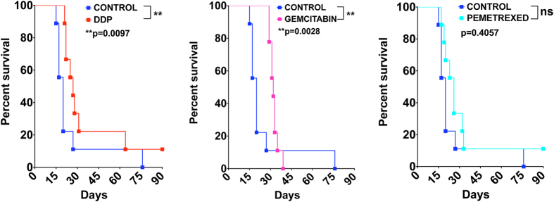
Malignant Mesothelioma is a highly aggressive cancer, which is difficult to diagnose and treat. Here we describe the molecular, cellular and morphological characterization of a syngeneic system consisting of murine AB1, AB12 and AB22 mesothelioma cells injected in immunocompetent BALB/c mice, which allows the study of the interplay of tumor cells with the immune system. Murine mesothelioma cells, like human ones, respond to exogenous High Mobility Group Box 1 protein, a Damage-Associated Molecular Pattern that acts as a chemoattractant for leukocytes and as a proinflammatory mediator. The tumors derived from AB cells are morphologically and histologically similar to human MM tumors, and respond to treatments used for MM patients. Our system largely recapitulates human mesothelioma, and we advocate its use for the study of MM development and treatment.


Figures

Figure 1
Figure 1. Morphology of murine MM cell lines.
Cultured AB1, AB12 and AB22 cells, showing morphological features corresponding to sarcomatoid (AB1; (a,d)), biphasic (AB12; (b,e)) and stellate/epithelioid (AB22; (c,f)) phenotypes. Boxed areas in a, b and c are enlarged in (df), respectively, to better appreciate cell morphology. Transmission electron microscopy reveals the presence of microvilli (g–i), a hallmark of mesothelial cells; Bars in g and h = 2 μm; bar in i = 4 μm.
Figure 2
Figure 2. Karyotype of murine MM cells.
Representative metaphases were visualized as described in Materials and Methods; 20 metaphases for each cell line were counted.
Figure 3
Figure 3. Murine MM cells express high levels of HMGB1 mRNA and secrete the protein.
(a) Total RNA from primary mesothelial cells was retrotranscribed and quantified by qPCR, as described in Materials and Methods. HMGB1 mRNA levels are about twofold higher in MM cell lines than in primary mesothelial cells (PMC). The bars represent standard deviation (n = 3). (b) Equal aliquots from total lysates of primary mesothelial cells or of the indicated cell lines were run (in duplicate) on SDS-PAGE, and western blotted with anti-HMGB1 and anti-β-actin antibodies. The filters were exposed for the same time showing that HMGB1 signal from primary mesothelial cells is barely detectable. (c) Quantification of HMGB1 bands in (b), normalized to β-actin levels. The bars represent standard deviation (n = 2).
Figure 4
Figure 4. Murine MM cells and tumors show both nuclear and cytoplasmic localization of HMGB1.
Nuclear and cytoplasmic localization of HMGB1 in cultured cells and tumor tissues. A polyclonal anti-HMGB1 antibody was used to immunostain (a) a cytopellet of cultured AB1-B/c-LUC cells, (b) a section of a tumor derived from them and (c) a section of a human MM. Bars = 100 μm.
Figure 5
Figure 5. AB cells respond to extracellular HMGB1.
(a) HMGB1 (30 ng/ml) acts as a chemoattractant for AB cell lines in Boyden chamber assays. The bars represent standard deviation (n = 3). (b) HMGB1 (30 ng/ml) increases the invasive potential of AB1-B/c-LUC and AB12-B/c-LUC cells, but not of AB22-B/c-LUC cells, in Boyden chamber invasion assays. The bars represent standard deviation (n = 3); p < 0.0001 (***). All experiments were repeated at least twice with similar results.
Figure 6
Figure 6. Tumor detection in vivo by BLI and US imaging.
15–20 days following injection of 7 × 104 AB1-B/c-LUC cells, mice developed tumor masses that were detected by BLI and US. The mass identified by US and shown in the panels on the right was estimated to measure 3.5 × 5.4 mm and yielded a higher BLI signal, whereas the one shown in the panels on the left was estimated to measure 2.5 × 2.5 mm and had a lower BLI signal. In both cases the BLI signal is sufficiently strong, allowing their detection as individual masses.
Figure 7
Figure 7. Vascularization of murine MM tumors.
Sarcomatoid tumors (red arrows) generated by injection of AB1 cells in BALB/c mice were explanted and stained with H&E (a,b); bar, 2 mm. No necrosis is detectable in the tumor masses. Immunostaining with anti-CD31 antibodies of sections from the tumor in (c) reveals several small vessels in the inner mass. (d) A movie montage (from Supplementary Movie 1) shows that the intravenously delivered contrast bolus infiltrates the tumor masses, demonstrating functional vascularization. (e,f) 3D (upper) and transaxial (lower) PET images of a mouse injected with AB1-B/c-LUC cells (f) show 18F-FDG signals (white arrows) in the abdomen, whereas images of a control animal do not (e). The higher signal in 3D PET images is due to the bladder, through which 18F-FDG is excreted. Images are presented with the same scale and were corrected for injected dose and animal weight. SUV: Standardized Uptake Value.
Figure 8
Figure 8. Murine and human tumors have similar morphologies.
Sections of explanted tumor masses generated by injection of AB1, AB12 and AB22 cells in BALB/c mice were stained with hematoxylin-eosin (H&E), as were sections from human sarcomatoid, biphasic and epithelioid mesotheliomas. The architecture of murine tumors appears similar to that of the corresponding human masses.
Figure 9
Figure 9. Immunohistochemical characterization of MM cell lines and tumors derived from them.
Following detachment from culture dishes, cells were fixed and centrifuged; the pellets were then sectioned and stained with the indicated antibodies (cytopellet). Explanted tumor masses were fixed, sectioned and stained with the same antibodies as in cytopellets. Arrows indicate positive cells. All pictures were taken with the same magnification (10X). Bars in top left panels of H&E stain of cytopellets and tumors = 50 μm.
Figure 10
Figure 10. Tumors generated by murine MM AB1-B/c-LUC cells respond to pharmacological treatments used for human MM.
Ten mice/group (control vs. treatment) were injected i.p. with 105 AB1-BALB/c-LUC cells. The Kaplan-Meier curves show that the treatment with DDP (a) significantly extends the survival of MM-bearing mice. Gemcitabin (b) significantly delays the onset of the disease. Pemetrexed (c) does not significantly affect the survival.

References

    1. Carbone M. et al. Malignant mesothelioma: facts, myths, and hypotheses. J Cell Physiol. 227, 44–58 (2012). - PMC - PubMed
    1. Carbone M. et al. BAP1 and cancer. Nat Rev Cancer 13, 153–159 (2013). - PMC - PubMed
    1. Husain A. N. et al. Guidelines for pathologic diagnosis of malignant mesothelioma: 2012 update of the consensus statement from the International Mesothelioma Interest Group. Arch Pathol Lab Med. 137, 647–667 (2013). - PubMed
    1. Rose A. H. et al. Increasing dietary selenium elevates reducing capacity and ERK activation associated with accelerated progression of select mesothelioma tumors. Am J Pathol. 184, 1041–1049 (2014). - PMC - PubMed
    1. Davidson B. Prognostic factors in malignant pleural mesothelioma. Hum Pathol. 46, 789–804 (2015). - PubMed

Publication types

MeSH terms