. 2016 Mar 9;36(10):2986-94.
doi: 10.1523/JNEUROSCI.2705-15.2016.

Pitch-Responsive Cortical Regions in Congenital Amusia

Affiliations

Pitch-Responsive Cortical Regions in Congenital Amusia

Sam V Norman-Haignere et al. J Neurosci. .

Abstract

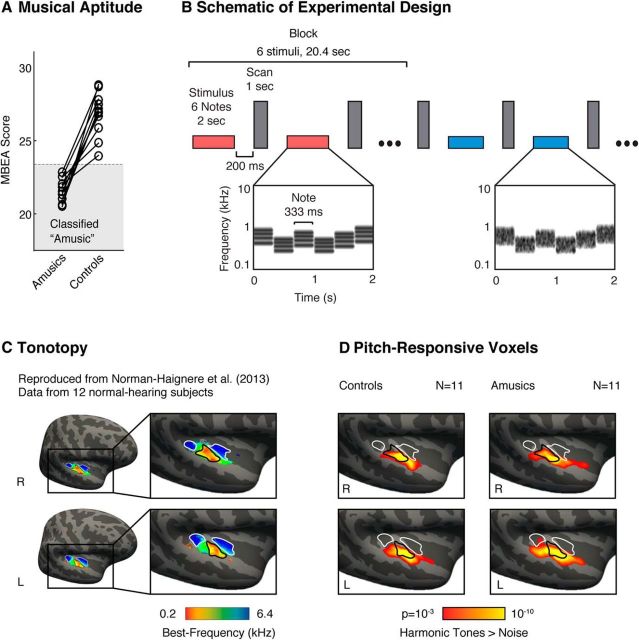
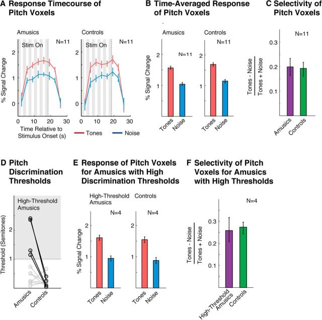
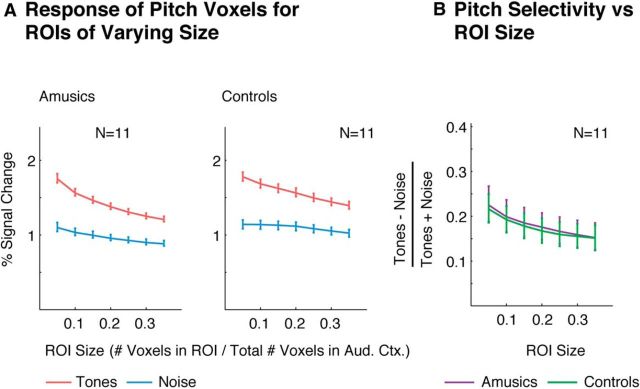
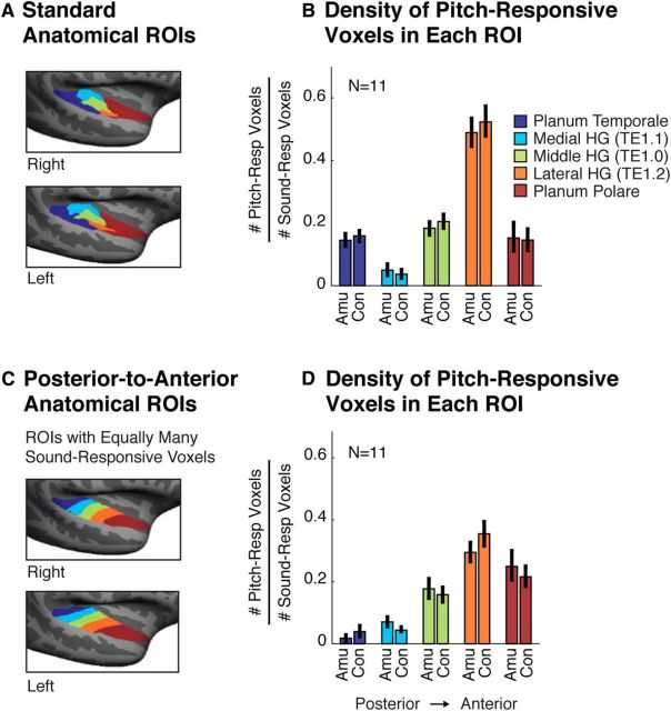
Congenital amusia is a lifelong deficit in music perception thought to reflect an underlying impairment in the perception and memory of pitch. The neural basis of amusic impairments is actively debated. Some prior studies have suggested that amusia stems from impaired connectivity between auditory and frontal cortex. However, it remains possible that impairments in pitch coding within auditory cortex also contribute to the disorder, in part because prior studies have not measured responses from the cortical regions most implicated in pitch perception in normal individuals. We addressed this question by measuring fMRI responses in 11 subjects with amusia and 11 age- and education-matched controls to a stimulus contrast that reliably identifies pitch-responsive regions in normal individuals: harmonic tones versus frequency-matched noise. Our findings demonstrate that amusic individuals with a substantial pitch perception deficit exhibit clusters of pitch-responsive voxels that are comparable in extent, selectivity, and anatomical location to those of control participants. We discuss possible explanations for why amusics might be impaired at perceiving pitch relations despite exhibiting normal fMRI responses to pitch in their auditory cortex: (1) individual neurons within the pitch-responsive region might exhibit abnormal tuning or temporal coding not detectable with fMRI, (2) anatomical tracts that link pitch-responsive regions to other brain areas (e.g., frontal cortex) might be altered, and (3) cortical regions outside of pitch-responsive cortex might be abnormal. The ability to identify pitch-responsive regions in individual amusic subjects will make it possible to ask more precise questions about their role in amusia in future work.

Keywords: amusia; auditory cortex; fMRI; music; pitch.


Figures

Figure 1.
Figure 1.
Schematic of experimental design and maps of pitch responses. A, Amusic subjects were identified using a standard musical aptitude test: the Montreal Battery of Evaluation of Amusia (MBEA). Each amusic subject was paired with a corresponding age- and education-matched control (matched pairs are indicated by connecting lines). B, Schematic of the experimental design. fMRI responses were measured to harmonic tones and Gaussian noise spanning the same frequency range. Contrasting responses to these stimuli in typical listeners reveals anatomically stereotyped “pitch-responsive” regions that overlap low- but not high-frequency tonotopic areas of primary auditory cortex (Norman-Haignere et al., 2013). Stimuli (denoted by horizontal bars) were presented in a block design, with six stimuli from the same condition presented successively in each block (red and blue indicate different conditions). Each stimulus (2 s) included several notes that varied in frequency to minimize adaptation. Cochleograms are shown for an example harmonic tone stimulus (red bar) and an example noise stimulus (blue bar). Cochleograms plot time–frequency decompositions, similar to a spectrogram, that summarize the cochlea's response to sound. After each stimulus, a single scan was collected (vertical, gray bars). C, Group tonotopic map reproduced from Norman-Haignere et al. (2013) and used here for comparison with pitch-responsive voxels (D). Colors indicate the pure-tone frequency with the highest response in each voxel. Black and white outlines indicate regions of low- and high-frequency selectivity, respectively. D, Voxels with a significant response preference for harmonic tones compared with frequency-matched noise (cluster-corrected to p < 0.001). In both amusics and controls, we observed significant clusters of pitch-responsive voxels that partially overlapped the low-frequency area of primary auditory cortex.
Figure 2.
Figure 2.
Pitch selectivity measured using functional ROIs. A, Response time course of pitch-responsive voxels to harmonic tones and noise, measured using an ROI analysis (see in “Functional ROI analyses” in the Materials and Methods). Gray regions indicate times when the stimulus was being played. The response to the stimulus is delayed because the BOLD response builds up slowly in response to neural activity. B, Time-averaged response to each condition (calculated using an HRF). C, Selectivity of the time-averaged response for harmonic tones compared with noise, measured by dividing the difference between responses to the two types of conditions by their sum. D, Pitch discrimination thresholds. Amusics' thresholds were higher on average than controls, but the amusic population was heterogeneous, consistent with prior reports (Tillmann et al., 2009; Liu et al., 2010). Four amusic subjects (bolded circles) had particularly high thresholds (>1 semitone) and were analyzed separately. E, Time-averaged response of pitch voxels for high-threshold amusics and their matched controls (same format as B). F, Selectivity of pitch-responsive voxels in high-threshold amusics and their matched controls (same format as C). Error bars in A, B, and E represent 1 SE of the mean difference between responses to harmonic tones and noise across subjects (computed via bootstrap); error bars in C and F represent 1 SE of the plotted selectivity measure across subjects (computed via bootstrap).
Figure 3.
Figure 3.
Extent of pitch selectivity measured using functional ROIs of varying size. A, Response of ROIs of varying size to harmonic tones and noise. Each ROI included the top N% of voxels in auditory cortex with the most significant response preference for harmonic tones compared with noise (selected using independent data from that used to measure the ROI's response). Error bars represent 1 SE of the mean difference between responses to harmonic tones and noise across subjects (computed via bootstrap). B, Selectivity of each ROI for harmonic tones compared with noise. Error bars represent 1 SE of the plotted selectivity measure across subjects (computed via bootstrap).
Figure 4.
Figure 4.
Anatomical distribution of pitch-responsive voxels. A, Five anatomical ROIs subdividing auditory cortex into standard regions (Morosan et al., 2001). Three ROIs subdivided Heschl's gyrus (HG) based on cytoarchitecture. Prior studies have reported a high density of pitch-responsive voxels in lateral HG (Patterson et al., 2002). The planum temporale and planum polare demarcate regions posterior and anterior to HG, respectively. B, Density of pitch-responsive voxels in each anatomical ROI, measured by the fraction of sound-responsive voxels (harmonic tones + noise > silence) that also exhibited a pitch response (harmonic tones > noise). C, Anatomical ROIs designed to run along the posterior-to-anterior axis of the superior temporal plane and to each include an equal number of sound-responsive voxels (Norman-Haignere et al., 2013). Prior work using these ROIs has shown a high density of pitch-responsive voxels in anterior regions of auditory cortex (Norman-Haignere et al., 2013). D, Density of pitch responses in each posterior-to-anterior ROI. Error bars in B and D represent 1 SE of the plotted density measure across subjects (computed via bootstrap).

References

    1. Albouy P, Mattout J, Bouet R, Maby E, Sanchez G, Aguera PE, Daligault S, Delpuech C, Bertrand O, Caclin A, Tillmann B. Impaired pitch perception and memory in congenital amusia: the deficit starts in the auditory cortex. Brain. 2013;136:1639–1661. doi: 10.1093/brain/awt082. - DOI - PubMed
    1. Albouy P, Mattout J, Sanchez G, Tillmann B, Caclin A. Altered retrieval of melodic information in congenital amusia: insights from dynamic causal modeling of MEG data. Front Hum Neurosci. 2015;9:20. doi: 10.3389/fnhum.2015.00020. - DOI - PMC - PubMed
    1. Angulo-Perkins A, Aubé W, Peretz I, Barrios FA, Armony JL, Concha L. Music listening engages specific cortical regions within the temporal lobes: differences between musicians and non-musicians. Cortex. 2014;59:126–137. doi: 10.1016/j.cortex.2014.07.013. - DOI - PubMed
    1. Avidan G, Tanzer M, Hadj-Bouziane F, Liu N, Ungerleider LG, Behrmann M. Selective dissociation between core and extended regions of the face processing network in congenital prosopagnosia. Cereb Cortex. 2014;24:1565–1578. doi: 10.1093/cercor/bht007. - DOI - PMC - PubMed
    1. Baldauf D, Desimone R. Neural mechanisms of object-based attention. Science. 2014;344:424–427. doi: 10.1126/science.1247003. - DOI - PubMed

Publication types

Supplementary concepts

LinkOut - more resources