. 2016 Aug;18(8):1088-1098.
doi: 10.1093/neuonc/now006. Epub 2016 Mar 8.

M011L-deficient oncolytic myxoma virus induces apoptosis in brain tumor-initiating cells and enhances survival in a novel immunocompetent mouse model of glioblastoma

Affiliations

M011L-deficient oncolytic myxoma virus induces apoptosis in brain tumor-initiating cells and enhances survival in a novel immunocompetent mouse model of glioblastoma

Alexandra Pisklakova et al. Neuro Oncol. 2016 Aug.

Abstract

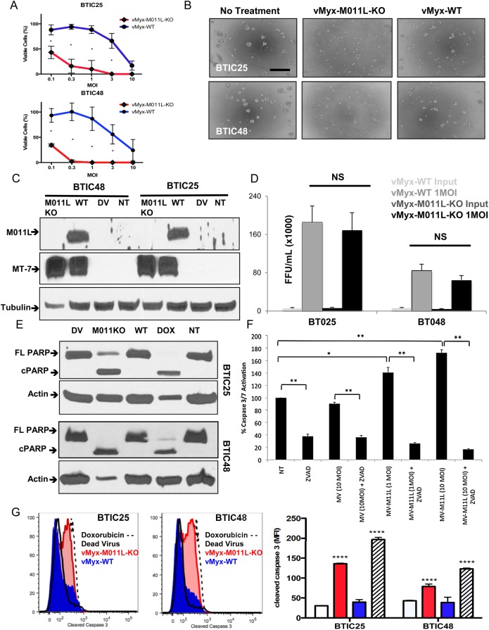
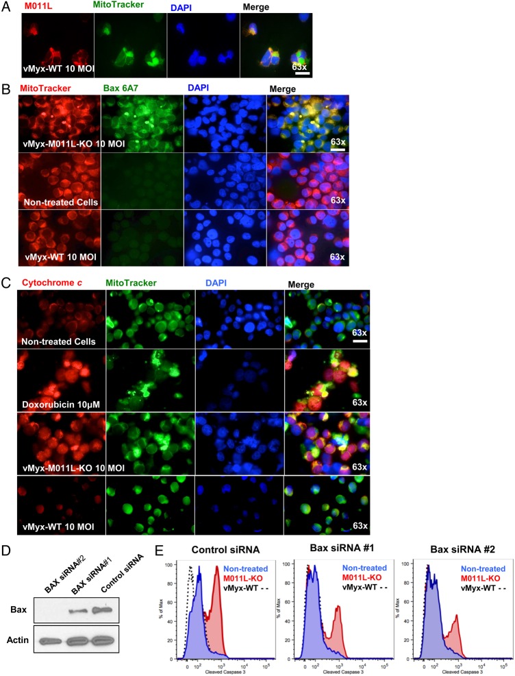
Background: Myxoma virus (MYXV) is a promising oncolytic agent and is highly effective against immortalized glioma cells but less effective against brain tumor initiating cells (BTICs), which are believed to mediate glioma development/recurrence. MYXV encodes various proteins to attenuate host cell apoptosis, including an antiapoptotic Bcl-2 homologue known as M011L. Such proteins may limit the ability of MYXV to kill BTICs, which have heightened resistance to apoptosis. We hypothesized that infecting BTICs with an M011L-deficient MYXV construct would overcome BTIC resistance to MYXV.

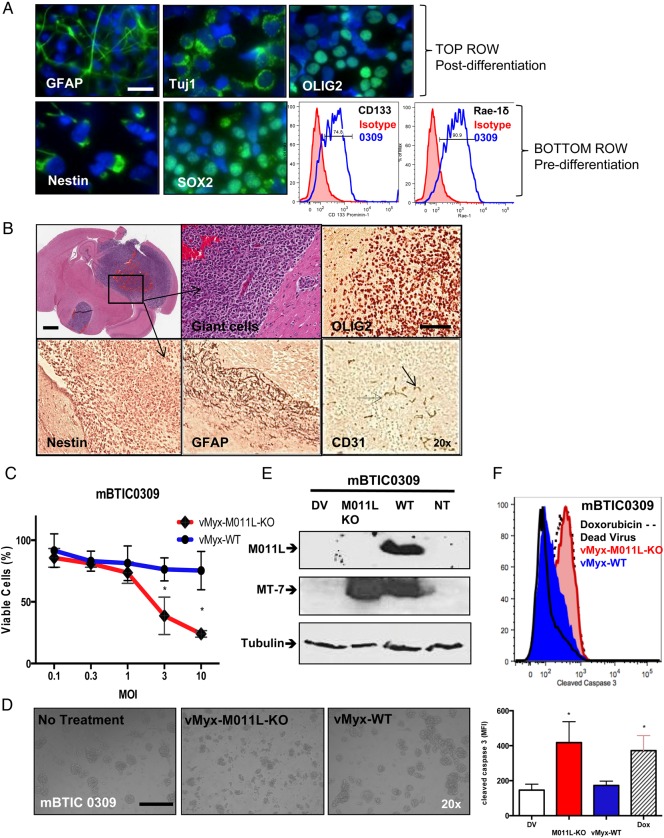
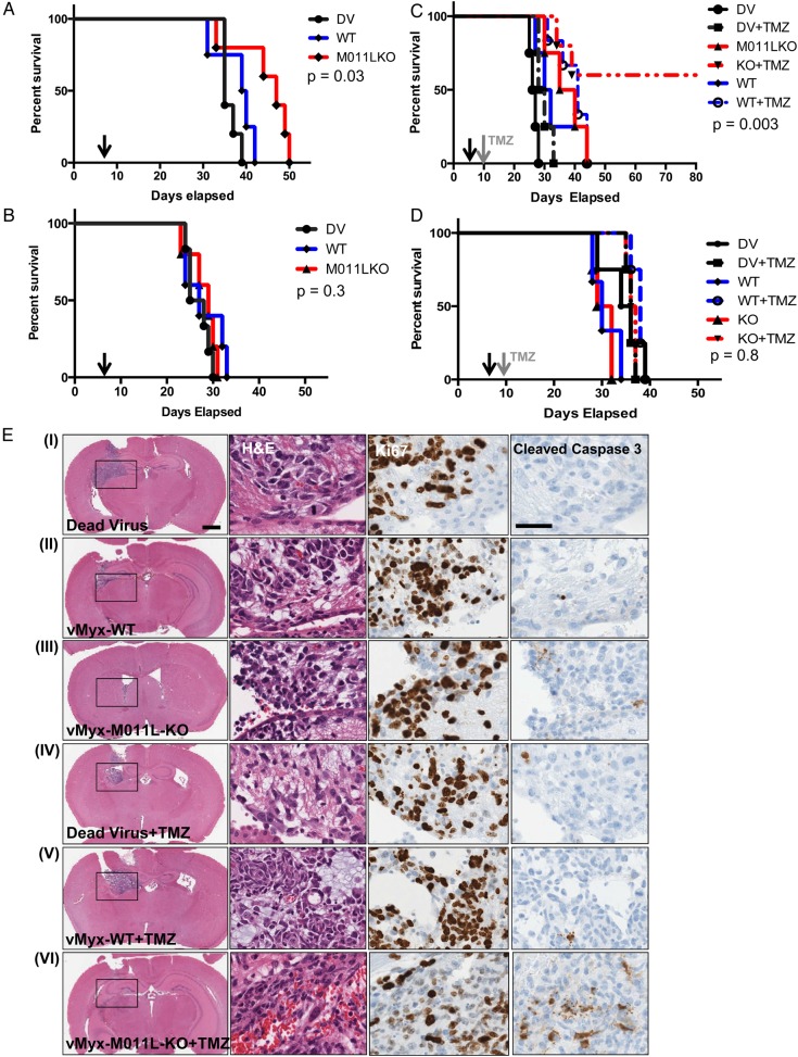
Methods: We used patient-derived BTICs to evaluate the efficacy of M011L knockout virus (vMyx-M011L-KO) versus wild-type MYXV (vMyx-WT) and characterized the mechanism of virus-induced cell death in vitro. To extend our findings in a novel immunocompetent animal model, we derived, cultured, and characterized a C57Bl/6J murine BTIC (mBTIC0309) from a spontaneous murine glioma and evaluated vMyx-M011L-KO efficacy with and without temozolomide (TMZ) in mBTIC0309-bearing mice.

Results: We demonstrated that vMyx-M011L-KO induces apoptosis in BTICs, dramatically increasing sensitivity to the virus. vMyx-WT failed to induce apoptosis as M011L protein prevented Bax activation and cytochrome c release. In vivo, intracranial implantation of mBTIC0309 generated tumors that closely recapitulated the pathological and molecular profile of human gliomas. Treatment of tumor-bearing mice with vMyx-M011L-KO significantly prolonged survival in immunocompetent-but not immunodeficient-mouse models, an effect that is significantly enhanced in combination with TMZ.

Conclusions: Our data suggest that vMyx-M011L-KO is an effective, well-tolerated, proapoptotic oncolytic virus and a strong candidate for clinical translation.

Keywords: apoptosis; brain tumor-initiating cells; glioma; oncolytic virus.


Figures

Fig. 1.
Fig. 1.
Infection with Myxoma virus M011L knockout virus (vMyx-M011L-KO), but not vMyx-wild-type (WT), induces apoptotic cell death in brain tumor-initiating cells (BTICs) in vitro. (A) Viability assay (Alamar Blue) of BTIC25 and BTIC48 72 hours post infection with vMyx-WT or vMyx-M011L-KO. vMyx-M011L-KO kills BTICs significantly more effectively than vMyx-WT in both BTICs tested. Data shown are mean ± SEM of experiments performed in triplicate, and results represent at least 3 independent experiments (*; P< .05 by Student t test). (B) vMyx-M011L-KO-treated BTICs show substantial cytopathic effects compared with vMyx-WT treated cells. Neurospheres were imaged using a Zeiss Axiovert microscope at 20× magnification, 96 hours post infection. Scale bar, 100 µm. Data are representative of 3 independent experiments. (C) BTIC25 and BTIC48 cells were infected with vMyx-WT (10 multiplicity of infection [MOI]), vMyx-M011L-KO (10 MOI), UV-inactivated MYXV (dead virus, DV) or left untreated (NT) for 24 hours prior to preparation of whole cell extracts. Extracts were immunoblotted for M011L, MT-7 (early viral gene expression), and tubulin (protein loading control). (D) The number of viral progeny produced by infected BTICs was quantified by counting the number of foci-forming units (FFUs) found on baby green monkey kidney (BGMK) cells treated with whole cell lysate from infected BTIC cultures. Titers were not significantly different between vMyx-M011L-KO and vMyx-WT in either BTIC25 or BTIC48. (E) PARP cleavage in BTICs induced by vMyx-M011L-KO. Cell extracts were prepared 48 hours post-infection with 10 MOI vMyx-M011L-KO, vMyx-WT, or UV-inactivated MYXV (DV) or doxorubicin treatment (10 μM; positive control). Full-length (FL) and cleaved (C) forms of poly ADP-ribose polymerase ( PARP) were assessed by immunoblotting using actin as a loading control. The figure shown is representative of results observed in 3 independent experiments. (F) Activation of caspase 3/7 in vMyx-WT and vMyx-M011L-KO-treated BTIC25 in vitro. Caspase 3/7 activation was assessed using Promega Caspase-Glo 3/7 assay. Luminescence is proportional to active caspase 3/7 in the sample, and was normalized to untreated BTIC25 (NT). Specificity of the assay for caspases was confirmed using the pan-caspase inhibitor ZVAD, which inhibited both constitutively active and vMyx-M011L-KO-activated caspases. vMyx-M011L-KO virus induced caspase 3/7 activation at both low (1 MOI) and high concentrations (10 MOI) of virus, whereas vMyx-WT (10 MOI) did not induce caspase 3/7 activation beyond the level observed in untreated (NT) cells. The data are representative of 3 independent experiments, and error bars represent mean ± SEM. *P < .05; **P < .01 by 2-sided t test. (G) BTICs were infected with vMyx-M011L-KO (10 MOI), vMyx-WT (10 MOI), UV-inactivated (dead) virus, or treated with doxorubicin (10 μM). Cells were collected after 72 hours, fixed, and stained with an antibody specific for cleaved caspase 3 and analyzed by flow cytometry. Data are representative of 3 independent experiments. Means ± SD from triplicates are shown (****P < .0001 by ANOVA compared with dead virus).
Fig. 2.
Fig. 2.
M011L protein localizes to mitochondria in brain-tumor initiating cells (BTICs) infected with vMyx-WT and prevents cytochrome c release by associating with proapoptotic protein Bax. (A) BTIC25 cells were infected with vMyx-WT (10 multiplicity of infection [MOI] for 24 hours prior to co-staining for M011L, mitochondria (Mitotracker Green FM) and nuclei (DAPI). Cells were visualized using an automated Zeiss Observer Z.1 inverted microscope through a 63X/1.4NA objective. M011L protein shows mitochondrial localization in cells infected with vMyx-WT. The data are representative of 3 independent experiments. (B) BTIC48 cells were infected with vMyx-WT (10 MOI), vMyx-M011L-KO (10 MOI), or left untreated for 6 hours prior to staining with an Alexa Fluor 488-labeled antibody to Bax (clone 6A7), Mitotracker Red, and DAPI. Note the co-localization (yellow) of active Bax with the mitochondria following infection with vMyx-M011L-KO (row 1, panel 4). Data represent 3 independent experiments. (C) BTIC48 were infected with vMyx-WT (10 MOI), vMyx-M011L-KO (10 MOI), treated with doxorubicin (10 μM) or left untreated for 10 hours prior to co-staining for cytochrome c (Alexa Fluor 546, red), mitochondria (MitoTracker Green FM), and nuclei (DAPI). Note increased staining for cytochrome c in cytosol (yellow) following treatment with doxorubicin (row 2, panel 4) and vMyx-M011L-KO (row 3, panel 4). Scale bar 25 µm. (D) BTIC48 cells were transfected with siRNAs specific for Bax or a control nontargeting pooled siRNA. After 48 hours, cells were collected, and Bax expression was determined by immunoblotting. (E) In parallel, 48 hours after transfection with siRNAs, BTICs were infected for 72 hours with either vMyx-M011L-KO (10 MOI), vMyx-WT (10 MOI), or left uninfected prior to assessing caspase 3 activation by flow cytometry.
Fig. 3.
Fig. 3.
Development and in vitro characterization of murine BTIC0309. (A) In vitro, neurospheres express neuronal markers SOX2, nestin, CD133 (Prominin-1) and Rae-1δ (NK-cell ligand, lower right fluorescence-activated cell sorting [FACS]) and can be differentiated in serum into glial (GFAP), neuronal (Tuj1), and oligodendrial (OLIG2) cell types. Scale bar 25 µm. (B) Mouse brain cross-section after intracranial implantation of mBTIC0309 at day 35 post infection. Scale bar 2 mm. Higher magnification of tumor tissue depicts cytonuclear pleomorphism and giant cells (upper middle). Tumors express endothelial cell marker CD31 and neuronal markers and maintain stem cell characteristics, as seen by immunohistochemistry of OLIG2, nestin and GFAP (brown staining) Scale bar 200 µm. (C) Alamar Blue assay of mBTIC0309 72 hours after infection with vMyx-WT or vMyx-M011L-KO. Data are mean ± SEM of experiments performed in triplicate, and results are representative of at least 3 independent experiments (*; P< .05 by Student t test). (D) vMyx-M011L-KO-treated mBTIC0309 shows substantial evidence of cytopathic effects compared with vMyx-WT treated cells. Neurospheres were imaged using a Zeiss Axiovert microscope at 20× magnification, 72 hours post infection. Data represent 3 independent experiments. (E) Immunoblot 24 hours after vMyx-WT or vMyxV-M011L-KO infection for M011L, MT-7 (early viral gene expression), and tubulin (protein loading control). (F) mBTIC0309 were infected with vMyx-M011L-KO (10 MOI), vMyx-WT (10 MOI), or UV-inactivated (dead) virus or treated with doxorubicin (10 μM). Cells were collected after 72 hours, fixed, and stained with an antibody for cleaved caspase 3 and analyzed by flow cytometry. Data are representative of 3 independent experiments. Means ± SD from triplicates are shown (*P< .05 by ANOVA compared with dead virus).
Fig. 4.
Fig. 4.
vMyx-M011L-KO prolongs survival of immunocompetent mice bearing syngeneic gliomas and results in long-term survival in combination with temozolomide (TMZ). Kaplan-Meier survival curve of mBTIC0309-bearing immunocompetent C57BL/6 mice (A) or immunodeficient NOD SCID gamma mice (B) treated with 5 × 10⁶ foci-forming units (FFUs) of vMyx-WT, vMyx-M011L-KO or UV-inactivated MYXV. Five mice were randomly assigned to each experimental group, and a single intratumoral virus injection was performed on day 7 after tumor implantation (black arrow). Statistical significance (P = .03, compared with vMyx-WT or dead virus, for C57BL/6J mice, (A) was determined using log-rank Mantel-Cox test. (C) Kaplan-Meier survival curve of mBTIC0309-bearing C57BL/6J mice treated with 5 × 10⁶ FFUs of vMyx-WT, vMyx-M011L-KO, or UV-inactivated MYXV in combination with TMZ. Ten mice were randomly assigned to each experimental group, and a single intratumoral virus injection was performed on day 7 after tumor implantation. Half of the animals were treated with 100 mg/kg of TMZ (grey arrow) on day 10 after tumor implantation (day 3 after virus treatment) for 5 consecutive days. Statistical significance (P < .007, vMyx-M011L-KO compared with dead virus, and P < .003, combination of TMZ and vMyx-M011L-KO compared with combination of TMZ and dead virus) was determined using log-rank Mantel-Cox test. No statistical significance (P = .8) in median survival was observed in immunocompromised animals treated in combination with TMZ (D). The data represent results from at least 2 independent experiments with similar results. (E) Immunohistochemical analysis 14 days after tumor inoculation (7 days after MYXV and 5 days after TMZ treatment). The tumor size was reduced in vMyx-M011L-KO + TMZ group (VI). Scale bar 2 mm. Combination treatment with vMyx-M011L-KO, and TMZ led to significantly (P < .05, Supplementary Material, Fig. S14) stronger cleaved caspase 3 staining, indicating increased apoptotic cell death in vivo. Scale bar 200 µm. Slides shown are representative of similar observations in 2 different mice receiving same treatment regimen.

Similar articles

Cited by

References

    1. Stupp R, Mason WP, van den Bent MJ et al. . Radiotherapy plus concomitant and adjuvant temozolomide for glioblastoma. N Engl J Med. 2005;352(10):987–996. - PubMed
    1. Singh SK, Hawkins C, Clarke ID et al. . Identification of human brain tumour initiating cells. Nature. 2004;432(7015):396–401. - PubMed
    1. Chen J, Li Y, Yu TS et al. . A restricted cell population propagates glioblastoma growth after chemotherapy. Nature. 2012;488(7412):522–526. - PMC - PubMed
    1. Kelly JJ, Stechishin O, Chojnacki A et al. . Proliferation of human glioblastoma stem cells occurs independently of exogenous mitogens. Stem Cells. 2009;27(8):1722–1733. - PubMed
    1. Lee J, Kotliarova S, Kotliarov Y et al. . Tumor stem cells derived from glioblastomas cultured in bFGF and EGF more closely mirror the phenotype and genotype of primary tumors than do serum-cultured cell lines. Cancer Cell. 2006;9(5):391–403. - PubMed