. 2016 Mar;13(116):20150932.
doi: 10.1098/rsif.2015.0932.

Analysis of blink dynamics in patients with blepharoptosis

Affiliations

Analysis of blink dynamics in patients with blepharoptosis

Felix H W Mak et al. J R Soc Interface. 2016 Mar.

Abstract

Owing to the rapid movements of the human upper eyelid, a high-speed camera was used to record and characterize voluntary blinking and the blink dynamics of blepharoptosis patients were compared to a control group. Twenty-six blepharoptosis patients prior to surgery and 45 control subjects were studied and the vertical height of the palpebral aperture (PA) was measured manually at 2 ms intervals during each blink cycle. The PA and blinking speed were plotted with respect to time and a predictive model was generated. The blink dynamic was analysed in closing and opening phases, and revealed a reduced speed of the initial opening phase in ptotic patients, suggesting intrinsic muscle function change in ptosis pathogenesis. The PA versus time curve for each subject was reconstructed using custom-built parameters; however, there were significant differences between the two groups. Those parameters used included the rate of closure, the delay between opening and closing, rate of initial opening, rate of slow opening (nonlinear function) and the 'switch point' between those two rates of opening. The model was tested against a new group of subjects and was able to discriminate ptosis patients from controls with 80% accuracy.

Keywords: blepharoptosis; blink dynamics; blinking; ptosis.


Figures

Figure 1.
Figure 1.
PA and speed master curves for all controls. Two sets of normal subject's blinking: Kwon et al. [13] PA master curve is shown in grey and its respective speed is shown in yellow [13]. The PA master curve is shown in blue and its respective speed is shown in orange. Secondary axis for the speed master curve is on the right-hand side, measured in millimetres per second. The chart is plotted with mean ± s.e.m.
Figure 2.
Figure 2.
PA and speed master curves for ptosis patients. PA versus speed master curve for both left and right eyes in ptosis patients. The PA master curve is shown in blue and its respective speed is shown in orange. Secondary axis for the speed master curve is on the right-hand side, measured in millimetres per second. The chart is plotted with mean ± s.e.m.
Figure 3.
Figure 3.
Fitting the blinking profile. An example of breaking down the blink PA versus time curve into possible key features—parameters that could be used in our analysis. The blue trace is the PA measurement against time, and the red trace is fitting of the functions designed to represent the key features of the blink profile.
Figure 4.
Figure 4.
Maximum speed during opening and closing phases: control versus ptosis (to the nearest number). The bars represents the averaged maximum speed during different phases of blinking: blue is control closing compared to orange for ptosis closing; and grey is control opening comparing to yellow for ptosis opening. Error bars are shown as s.e.m.
Figure 5.
Figure 5.
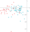
Blinking discriminant between control subjects of greater than or equal to 40 (pink) versus less than 40 (cyan). This analysis used the five parameters mentioned in the text: rate of closure, delay between opening and closing, initial rate of opening, switch point and rate of slow opening. The coloured X is the mean of the respective group. Values on x1- and x2-axes are merely numbers representing the reduced dimension in LDA.
Figure 6.
Figure 6.
Three-dimensional discriminant display of the parameter used for ptosis (all ages; green) and controls (greater than or equal to 40 year-old; orange). v1, v2, topen and tclose are defined in text (§2.5). The value of PAmin is neglected as it does not have much significance, as the real value of the minimum is often at a cusp. Also the start and end % were not considered because they are not really shape parameters.
Figure 7.
Figure 7.
Ptosis patients (green) versus greater than or equal to age 40 controls (orange) in the reduced dimension of the linear discriminant. Axes x1 and x2 are the first two principal directions in the space of the linear discriminant. The class means are shown as the X marker in its respective colour.
Figure 8.
Figure 8.
Comparison of v2 parameter between ptosis and control patients are displayed on this histogram. It demonstrates a slower rate of opening in the ptosis patients than the controls by a small, but significant amount.
Figure 9.
Figure 9.
Fitting of the blinking profile on the PA versus time curve for a control patient. It is clear that the blinking profile is not fitting every detail if we only have linear functions for closure and initial opening. Some of the information is lost after conversion into the fitting function.

References

    1. Sudhakar P, Vu Q, Kosoko-Lasaki O, Palmer M. 2009. Upper eyelid ptosis revisited. Am. J. Clin. Med. 6, 5–14.
    1. Le TT, Farkas LG, Ngim RCK, Levin LS, Forrest CR. 2002. Proportionality in Asian and North American Caucasian faces using neoclassical facial canons as criteria. Aesthetic Plastic Surgery 26, 64–69. (10.1007/s00266-001-0033-7) - DOI - PubMed
    1. Dortzbach RK, Sutula FC. 1980. Involutional belpharoptosis. A histological study. Arch. Ophthalmol. 98, 2045–2049. (10.1001/archopht.1980.01020040897022) - DOI - PubMed
    1. Fujiwara T, Matsuo K, Kondoh S, Yuzuriha S. 2001. Etiology and pathogenesis of aponeurotic blepharoptosis. Ann. Plast. Surg. 46, 29–35. (10.1097/00000637-200101000-00006) - DOI - PubMed
    1. Paris GL, Quickert MH. 1976. Disinsertion of the aponeurosis of the levator palpebrae superioris muscle after cataract extraction. Am. J. Ophthalmol. 81, 337–340. (10.1016/0002-9394(76)90250-6) - DOI - PubMed

Publication types

LinkOut - more resources