. 2016 Apr 15;5(4):409-17.
doi: 10.1242/bio.015404.

Cross-species functional analyses reveal shared and separate roles for Sox11 in frog primary neurogenesis and mouse cortical neuronal differentiation

Affiliations

Cross-species functional analyses reveal shared and separate roles for Sox11 in frog primary neurogenesis and mouse cortical neuronal differentiation

Chao Chen et al. Biol Open. .

Abstract

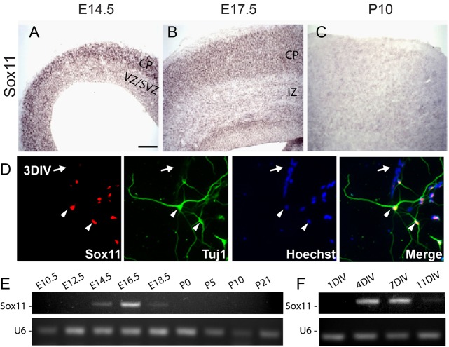
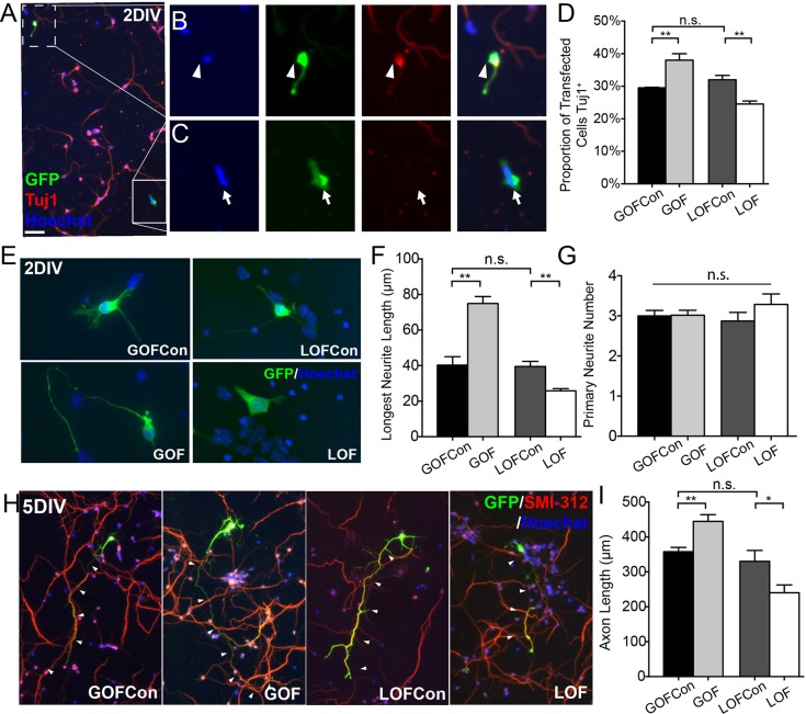
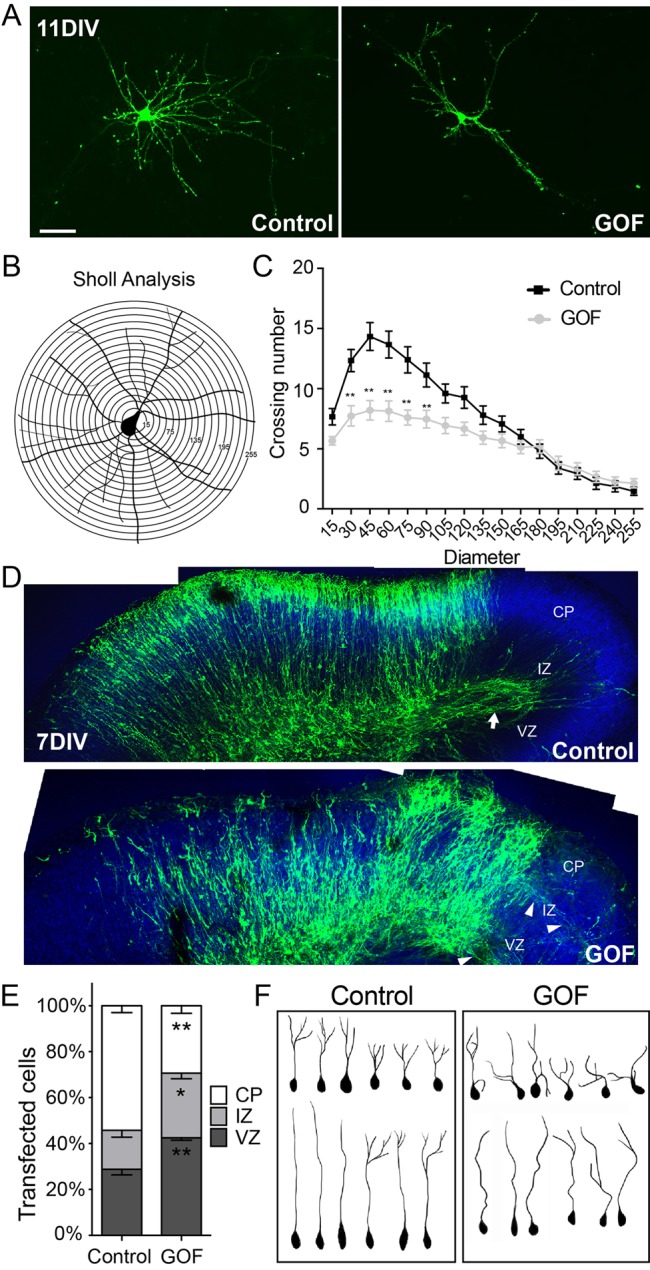
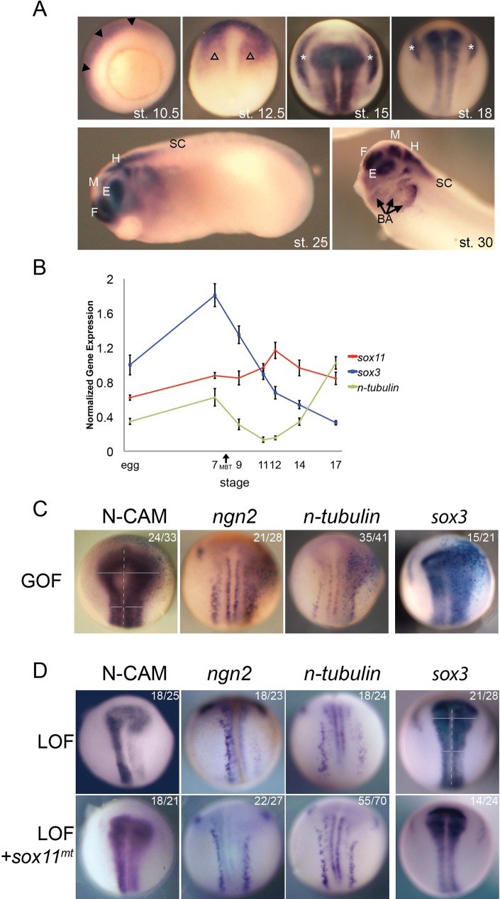
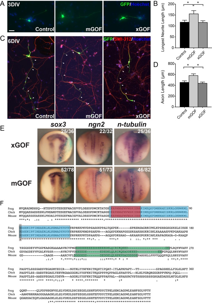
A well-functioning brain requires production of the correct number and types of cells during development; cascades of transcription factors are essential for cellular coordination. Sox proteins are transcription factors that affect various processes in the development of the nervous system. Sox11, a member of the SoxC family, is expressed in differentiated neurons and supports neuronal differentiation in several systems. To understand how generalizable the actions of Sox11 are across phylogeny, its function in the development of the frog nervous system and the mouse cerebral cortex were compared. Expression of Sox11 is largely conserved between these species; in the developing frog, Sox11 is expressed in the neural plate, neural tube and throughout the segmented brain, while in the mouse cerebral cortex, Sox11 is expressed in differentiated zones, including the preplate, subplate, marginal zone and cortical plate. In both frog and mouse, data demonstrate that Sox11 supports a role in promoting neuronal differentiation, with Sox11-positive cells expressing pan-neural markers and becoming morphologically complex. However, frog and mouse Sox11 cannot substitute for one another; a functional difference likely reflected in sequence divergence. Thus, Sox11 appears to act similarly in subserving neuronal differentiation but is species-specific in frog neural development and mouse corticogenesis.

Keywords: Neural development; Neuronal differentiation; Sox transcription factor.


Conflict of interest statement

Competing interests

The authors declare no competing or financial interests.

Figures

Fig. 1.
Fig. 1.
Sox11 is expressed in differentiated neurons in vivo and in vitro and is dynamically expressed during mouse corticogenesis. (A-C) Wild type cerebral cortex hybridized with antisense probes specific for Sox11 at E14.5 (A), E17.5 (B) and P10 (C), with expression visualized using BM Purple. Sox11 is expressed in the cortical plate (CP) at E14.5 and E17.5. There is no detectable expression of Sox11 at P10. (D) In cultured cortical neurons examined after 3 days in vitro (3DIV), expression of Sox11 is localized to cells that are TuJ1+ (arrowheads) and absent from TuJ1 cells (arrow). (E-F) Profile of Sox11 expression during cortical development in vivo (E) and in vitro (F) reveals undetectable levels early (E10.5-E12.5 and 1DIV), activation (E14.5-E18.5 and 4DIV), and inactivation (P0-P21 and 11DIV). CP, cortical plate; VZ, ventricular zone; SVZ, sub-ventricular zone; IZ, intermediate zone. Scale bar: 60 μm (A,B); 130 μm (C); 25 μm (D).
Fig. 2.
Fig. 2.
Perturbation of Sox11 shifts proportions of neurons and alters neuronal morphology of mouse cortical neurons in vitro. (A-C) A representative image of cortical neurons transfected with GFP (green), grown for 2DIV, and stained with TuJ1 (red) and Hoechst (blue) (A). GFP+-transfected cells are either TuJ1+ neurons (B, arrowhead) or TuJ1 cells (C, arrow). (D) The proportion of transfected cells that are TuJ1+ is greater in Sox11GOF and lower in Sox11LOF, compared to appropriate control cultures. (E-G) Cells transfected with control (top), Sox11GOF (bottom left), or Sox11LOF (bottom right), stained for GFP and labeled with Hoechst, and imaged at 2DIV (E). The average length of the longest neurite is longer in Sox11GOF and shorter in Sox11LOF compared to controls (F) whereas there is no difference in the number of primary neurites (G). (H-I) GFP+ cells transfected with control vectors (left and third from left), Sox11GOF (second from left), or Sox11LOF (right) were harvested at 5 DIV and stained with SMI-312 (red) to mark axons (H). Average axon length was longer in Sox11GOF and shorter in Sox11LOF compared to control cells (I). Scale bar: 60 μm (A); 24 μm (B,C); 12 μm (E); 52 μm (I). Data represented as mean±sem; n.s., not significant; *P<0.05; **P<0.01.
Fig. 3.
Fig. 3.
Misexpression of Sox11 in mouse cortical neurons interferes with dendritic extent in vitro and localization and shape of neurons in vivo. (A-C) Images of primary cortical neurons expressing GFP alone (left) or GFP and Sox11 (right) (A). A schematic of Sholl analysis of a cortical neuron in which neurite crossings for each ring are quantified (B). Overexpression of Sox11 (gray) reduced the complexity of dendritic branching of cortical neurons in vitro compared to control neurons (black) (C). (D) Organotypic slice cultures of cortex transfected at E14.5 with GFP alone (top) or GFP and Sox11 (bottom) and imaged at 7DIV. GFP+ cells have extended an axon tract in control (arrow) while in Sox11GOF, axons were diffuse (arrowheads). (E) Distribution of GFP+ cells in cortical embryonic zones is different in Sox11GOF compared to control; Sox11GOF cells tend to remain closer to the lateral ventricle, in the ventricular zone (VZ) and intermediate zone (IZ), than control-transfected cells. (F) Representative traces of neurons from control (left) and Sox11GOF (right) cortical plate (CP) reveals that neurons are less orderly when Sox11 expression is maintained. Scale bar: 30 μm (A); 63 μm (D). Data represented as mean±sem; *P<0.05; **P<0.01.
Fig. 4.
Fig. 4.
Sox11 is expressed in neural tissue and influences neural development in Xenopus laevis. (A) Whole mount in situ hybridization (WISH) of sox11 in frog embryos at st. 10.5 (vegetal view), 12.5, 15 and 18 (dorsal view along anterior-posterior axis), 25 (lateral view, with head to the left), 30 (lateral view of head) reveals sox11 is expressed in the presumptive CNS. Filled arrowhead marks ectoderm. Open arrowhead marks anterior neural plate. Asterisk marks intermediate and posterior placodes. Arrow marks branchial arches. F, forebrain; M, midbrain; H, hindbrain; E, eye; SC, spinal cord; BA, branchial arches. (B) Quantitative PCR of sox3, n-tubulin and sox11 expression during frog development reveals sox11 is expressed throughout early embryonic development. Data represented as mean±sem. (C-D) WISH of st. 15 embryos injected on the right side with sox11 RNA (dorsal view, along the anterior-posterior axis). sox11GOF expands expression of pan-neural marker, N-CAM, the proneural gene, ngn2, the neuronal marker, n-tubulin, and the neural progenitor marker, sox3. (D) WISH of st. 15 embryos injected on the right side with Sox11 morpholinos alone (top) or Sox11 morpholinos and sox11mt RNA (bottom). When Sox11 is reduced, expressions of N-CAM, ngn2 and n-tubulin were decreased, and expression of sox3 was increased; all LOF phenotypes were reversed when sox11mt was expressed.
Fig. 5.
Fig. 5.
Functional reciprocity of Sox11 between frog and mouse. (A-D) Primary cortical neurons expressing GFP alone (left), GFP and mouse Sox11 (mGOF, middle), and GFP and Xenopus sox11 (xGOF, right) and analyzed at 3DIV (A,B) and 6DIV (C,D) revealed increases in the longest neurite (B) and axon (D) when mouse Sox11 but not frog sox11 was expressed. Data represented as mean±sem; *P<0.05. (E) WISH of st. 15 embryos (dorsal view, along with the anterior-posterior axis) injected at the two-cell stage with either frog (top) or mouse (bottom) sox11 and lacZ tracer (blue). xGOF elevated levels of sox3, ngn2 and n-tubulin, while mGOF had no effect. (F) Alignment of frog, chicken and mouse Sox11 protein sequences. Frog sequence shown here is Sox11a sequence: Sox11a and Sox11b share 93% similarity, and the single amino acid difference observed between frog Sox11a and other species is also present in Sox11b sequence. Discrete protein domains are shaded: blue is the HMG box, red is the nuclear localization signal; green is the acid-rich region; grey marks the one amino acid difference in the HMG box. (*=Identical amino acid residues; :=Different but highly conserved amino acid residues;∙=Different but somewhat similar amino acid residues; blank=dissimilar amino acid residues or gap.) Scale bar: 32 μm (A); 84 μm (C).

References

    1. Amin N. M., Tandon P., Osborne Nishimura E. and Conlon F. L. (2014). RNA-seq in the tetraploid Xenopus laevis enables genome-wide insight in a classic developmental biology model organism. Methods 66, 398-409. 10.1016/j.ymeth.2013.06.009 - DOI - PMC - PubMed
    1. Bergsland M., Werme M., Malewicz M., Perlmann T. and Muhr J. (2006). The establishment of neuronal properties is controlled by Sox4 and Sox11. Genes Dev. 20, 3475-3486. 10.1101/gad.403406 - DOI - PMC - PubMed
    1. Bergsland M., Ramskold D., Zaouter C., Klum S., Sandberg R. and Muhr J. (2011). Sequentially acting Sox transcription factors in neural lineage development. Genes Dev. 25, 2453-2464. 10.1101/gad.176008.111 - DOI - PMC - PubMed
    1. Bowles J., Schepers G. and Koopman P. (2000). Phylogeny of the SOX family of developmental transcription factors based on sequence and structural indicators. Dev. Biol. 227, 239-255. 10.1006/dbio.2000.9883 - DOI - PubMed
    1. Braman J., Papworth C. and Greener A. (1996). Site-directed mutagenesis using double-stranded plasmid DNA templates. Methods Mol. Biol. 57, 31-44. 10.1385/0-89603-332-5:31 - DOI - PubMed