Primary Retroperitoneal Mucinous Cystadenoma
- PMID: 26962534
- PMCID: PMC4783510
- DOI: 10.3393/ac.2016.32.1.33
Primary Retroperitoneal Mucinous Cystadenoma
Abstract
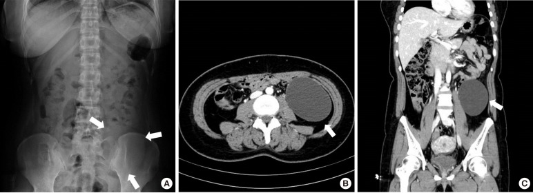
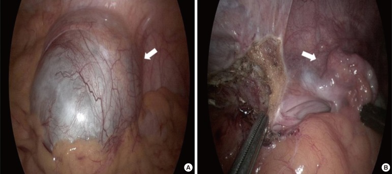
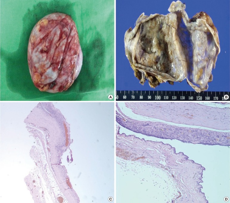
Mucinous cystadenomas and cystadenocarcinomas of the ovary are clinically and histopathologically well-established common tumors. However, primary retroperitoneal mucinous cystic tumors are extremely rare, and although their histopathogenesis is still uncertain, several theories have been proposed. Most authors suggest that they develop through mucinous metaplasia in a preexisting mesothelium-lined cyst. An accurate preoperative diagnosis of these tumors is difficult because no effective diagnostic measures have been established. Delay in diagnosis and treatment of this tumor may be fatal for the patient because of complications such as rupture, infection, and malignant transformation. We describe the case of a 31-year-old woman with abdominal pain and a palpable mass. Computed tomography of the abdomen revealed a retroperitoneal cystic mass, which was resected successfully through laparoscopy. Histopathological examination of the resected mass confirmed the diagnosis of a primary retroperitoneal mucinous cystadenoma. The patient was discharged on postoperative day 5 without any complications.
Keywords: Mucinous cystadenoma; Retroperitoneal neoplasms.
Conflict of interest statement
Figures
References
-
- Lai EC, Chung KM, Lau WY. Primary retroperitoneal mucinous cystadenoma. ANZ J Surg. 2006;76:537. - PubMed
-
- Arribas D, Cay A, Latorre A, Cordoba E, Martinez F, Lagos J. Retroperitoneal mucinous cystadenoma. Arch Gynecol Obstet. 2004;270:292–293. - PubMed
-
- Subramony C, Habibpour S, Hashimoto LA. Retroperitoneal mucinous cystadenoma. Arch Pathol Lab Med. 2001;125:691–694. - PubMed
LinkOut - more resources
Full Text Sources
Other Literature Sources