

Overcoming MITF-conferred drug resistance through dual AURKA/MAPK targeting in human melanoma cells
- PMID: 26962685
- PMCID: PMC4823922
- DOI: 10.1038/cddis.2015.369
Overcoming MITF-conferred drug resistance through dual AURKA/MAPK targeting in human melanoma cells
Abstract
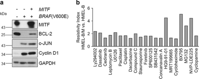
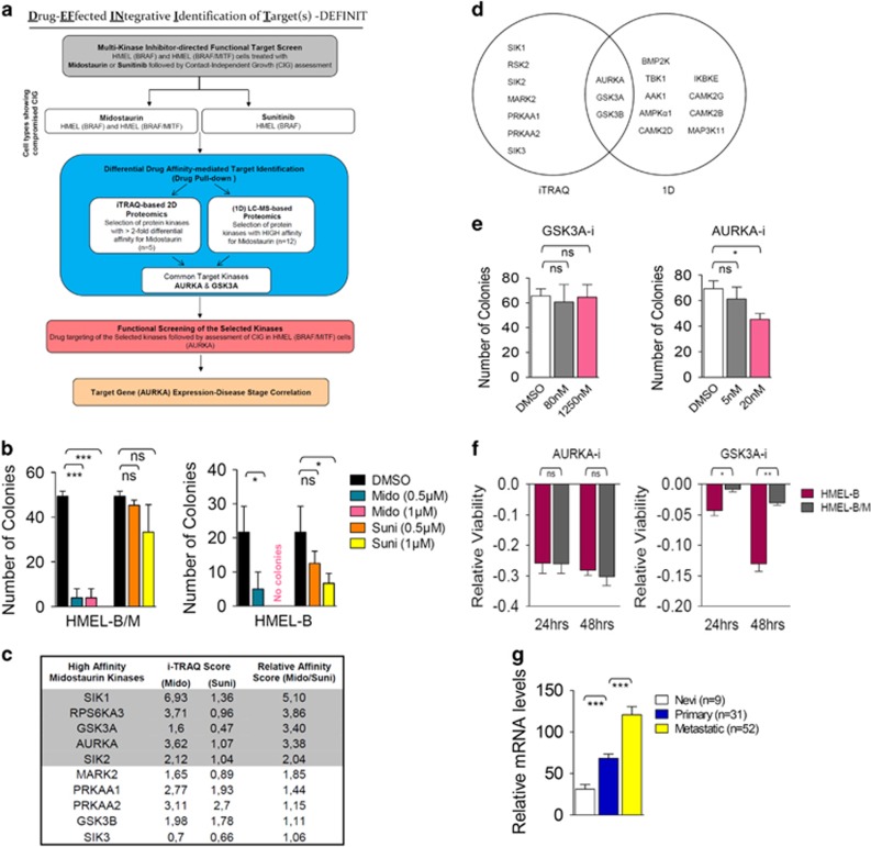
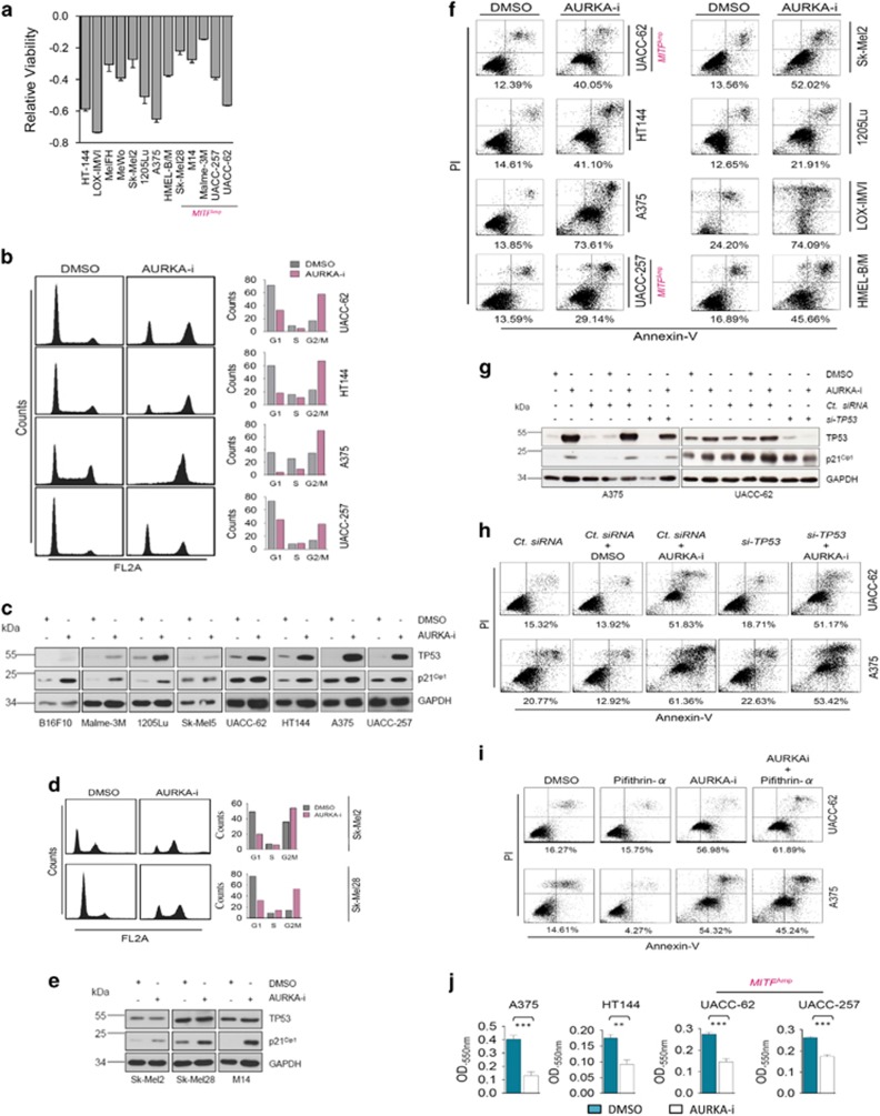
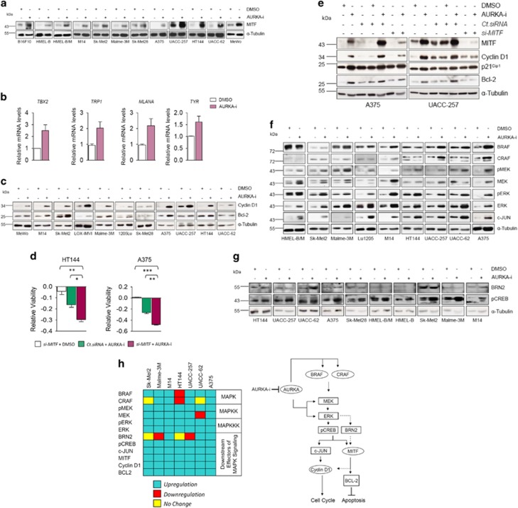
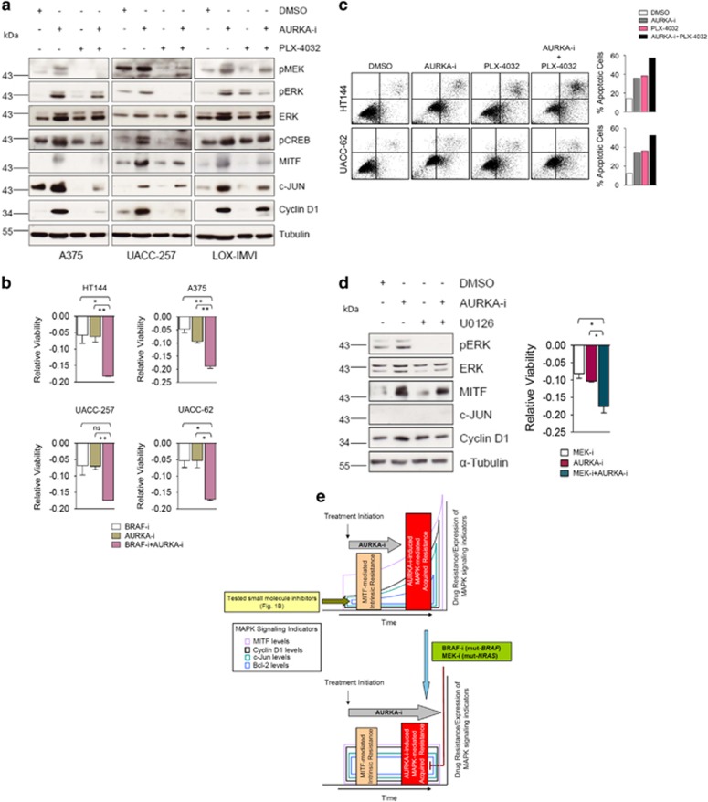
MITF (microphthalmia-associated transcription factor) is a frequently amplified lineage-specific oncogene in human melanoma, whose role in intrinsic drug resistance has not been systematically investigated. Utilizing chemical inhibitors for major signaling pathways/cellular processes, we witness MITF as an elicitor of intrinsic drug resistance. To search kinase(s) targets able to bypass MITF-conferred drug resistance, we employed a multi-kinase inhibitor-directed chemical proteomics-based differential affinity screen in human melanocytes carrying ectopic MITF overexpression. A subsequent methodical interrogation informed mitotic Ser/Thr kinase Aurora Kinase A (AURKA) as a crucial regulator of melanoma cell proliferation and migration, independent of the underlying molecular alterations, including TP53 functional status and MITF levels. Crucially, assessing the efficacy of investigational AURKA inhibitor MLN8237, we pre-emptively witness the procurement of a molecular program consistent with acquired drug resistance. This involved induction of multiple MAPK (mitogen-activated protein kinase) signaling pathway components and their downstream proliferation effectors (Cyclin D1 and c-JUN) and apoptotic regulators (MITF and Bcl-2). A concomitant AURKA/BRAF and AURKA/MEK targeting overcame MAPK signaling activation-associated resistance signature in BRAF- and NRAS-mutated melanomas, respectively, and elicited heightened anti-proliferative activity and apoptotic cell death. These findings reveal a previously unreported MAPK signaling-mediated mechanism of immediate resistance to AURKA inhibitors. These findings could bear significant implications for the application and the success of anti-AURKA approaches that have already entered phase-II clinical trials for human melanoma.
Figures
Similar articles
-
P53 and MITF/Bcl-2 identified as key pathways in the acquired resistance of NRAS-mutant melanoma to MEK inhibition.Eur J Cancer. 2017 Sep;83:154-165. doi: 10.1016/j.ejca.2017.06.033. Epub 2017 Jul 21. Eur J Cancer. 2017. PMID: 28738256
-
Mitogen-activated protein kinase (MAPK) hyperactivation and enhanced NRAS expression drive acquired vemurafenib resistance in V600E BRAF melanoma cells.J Biol Chem. 2014 Oct 3;289(40):27714-26. doi: 10.1074/jbc.M113.532432. Epub 2014 Jul 25. J Biol Chem. 2014. PMID: 25063807 Free PMC article.
-
AurkA inhibitors enhance the effects of B-RAF and MEK inhibitors in melanoma treatment.J Transl Med. 2014 Jul 31;12:216. doi: 10.1186/s12967-014-0216-z. J Transl Med. 2014. PMID: 25074438 Free PMC article.
-
Novel mechanisms and therapeutic approaches in melanoma: targeting the MAPK pathway.Discov Med. 2015 Jun;19(107):455-61. Discov Med. 2015. PMID: 26175403 Review.
-
MAPK pathway inhibition in melanoma: resistance three ways.Biochem Soc Trans. 2014 Aug;42(4):727-32. doi: 10.1042/BST20140020. Biochem Soc Trans. 2014. PMID: 25109949 Review.
Cited by
-
Pueraria protein extract inhibits melanogenesis and promotes melanoma cell apoptosis through the regulation of MITF and mitochondrial‑related pathways.Mol Med Rep. 2023 Mar;27(3):64. doi: 10.3892/mmr.2023.12951. Epub 2023 Feb 3. Mol Med Rep. 2023. PMID: 36734267 Free PMC article.
-
Cuprous oxide nanoparticle-inhibited melanoma progress by targeting melanoma stem cells.Int J Nanomedicine. 2017 Apr 5;12:2553-2567. doi: 10.2147/IJN.S130753. eCollection 2017. Int J Nanomedicine. 2017. PMID: 28435246 Free PMC article.
-
Network Pharmacology Integrated Molecular Docking Reveals the Antiosteosarcoma Mechanism of Biochanin A.Evid Based Complement Alternat Med. 2019 Jan 6;2019:1410495. doi: 10.1155/2019/1410495. eCollection 2019. Evid Based Complement Alternat Med. 2019. PMID: 30723510 Free PMC article.
-
Identification of New Vulnerabilities in Conjunctival Melanoma Using Image-Based High Content Drug Screening.Cancers (Basel). 2022 Mar 19;14(6):1575. doi: 10.3390/cancers14061575. Cancers (Basel). 2022. PMID: 35326726 Free PMC article.
-
Molecular Targeting of HuR Oncoprotein Suppresses MITF and Induces Apoptosis in Melanoma Cells.Cancers (Basel). 2021 Jan 6;13(2):166. doi: 10.3390/cancers13020166. Cancers (Basel). 2021. PMID: 33418925 Free PMC article.
References
-
- Davies H, Bignell GR, Cox C, Stephens P, Edkins S, Clegg S et al. Mutations of the BRAF gene in human cancer. Nature 2002; 417: 949–954. - PubMed
-
- Jang S, Atkins MB. Treatment of BRAF-mutant melanoma: the role of vemurafenib and other therapies. Clin Pharmacol Ther 2014; 95: 24–31. - PubMed
-
- Sun C, Wang L, Huang S, Heynen GJ, Prahallad A, Robert C et al. Reversible and adaptive resistance to BRAF(V600E) inhibition in melanoma. Nature 2014; 508: 118–122. - PubMed
Publication types
MeSH terms
Substances
LinkOut - more resources
Full Text Sources
Other Literature Sources
Medical
Research Materials
Miscellaneous
