

Can a total knee arthroplasty be both rotationally unconstrained and anteroposteriorly stabilised? A pulsed fluoroscopic investigation
- PMID: 26965166
- PMCID: PMC4852793
- DOI: 10.1302/2046-3758.53.2000621
Can a total knee arthroplasty be both rotationally unconstrained and anteroposteriorly stabilised? A pulsed fluoroscopic investigation
Abstract
Objectives: Throughout the 20th Century, it has been postulated that the knee moves on the basis of a four-bar link mechanism composed of the cruciate ligaments, the femur and the tibia. As a consequence, the femur has been thought to roll back with flexion, and total knee arthroplasty (TKA) prostheses have been designed on this basis. Recent work, however, has proposed that at a position of between 0° and 120° the medial femoral condyle does not move anteroposteriorly whereas the lateral femoral condyle tends, but is not obliged, to roll back - a combination of movements which equates to tibial internal/ femoral external rotation with flexion. The aim of this paper was to assess if the articular geometry of the GMK Sphere TKA could recreate the natural knee movements in situ/in vivo.
Methods: The pattern of knee movement was studied in 15 patients (six male: nine female; one male with bilateral TKAs) with 16 GMK Sphere implants, at a mean age of 66 years (53 to 76) with a mean BMI of 30 kg/m(2) (20 to 35). The motions of all 16 knees were observed using pulsed fluoroscopy during a number of weight-bearing and non-weight-bearing static and dynamic activities.
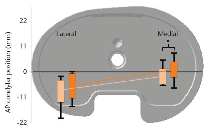
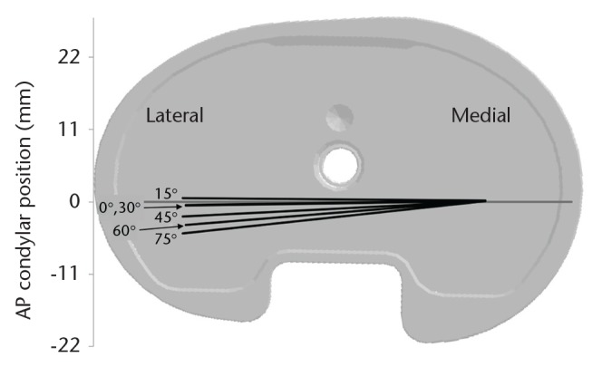
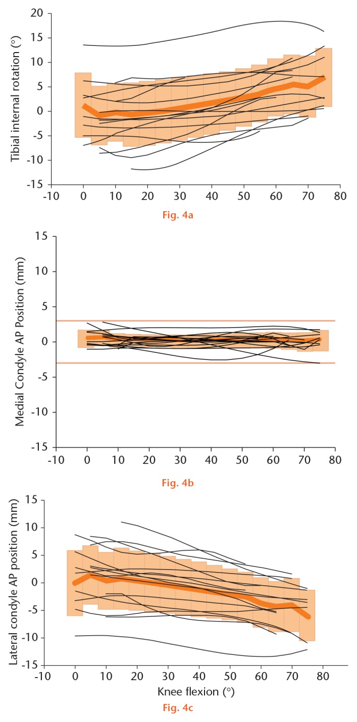
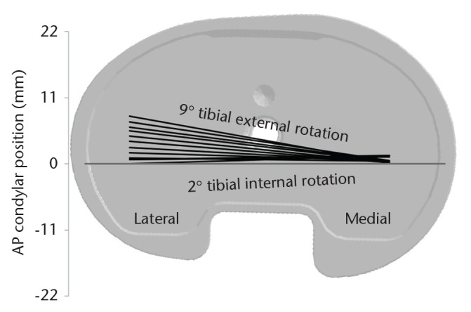
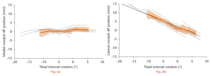
Results: During maximally flexed kneeling and lunging activities, the mean tibial internal rotation was 8° (standard deviation (sd) 6). At a mean 112° flexion (sd 16) during lunging, the medial and lateral condyles were a mean of 2 mm (sd 3) and 8 mm (sd 4) posterior to a transverse line passing through the centre of the medial tibial concavity. With a mean flexion of 117° (sd 14) during kneeling, the medial and lateral condyles were a mean of 1 mm (sd 4) anterior and 6 mm (sd 4) posterior to the same line. During dynamic stair and pivoting activities, there was a mean anteroposterior translation of 0 mm to 2 mm of the medial femoral condyle. Backward lateral condylar translation occurred and was linearly related to tibial rotation.
Conclusion: The GMK Sphere TKA in our study group shows movements similar in pattern, although reduced in magnitude, to those in recent reports relating to normal knees during several activities. Specifically, little or no translation of the medial femoral condyle was observed during flexion, but there was posterior roll-back of the lateral femoral condyle, equating to tibiofemoral rotation. We conclude that the GMK Sphere is anteroposteriorly stable medially and permits rotation about the medial compartment.Cite this article: Professor G. Scott. Can a total knee arthroplasty be both rotationally unconstrained and anteroposteriorly stabilised?: A pulsed fluoroscopic investigation. Bone Joint Res 2016;5:80-86. DOI: 10.1302/2046-3758.53.2000621.
Keywords: TKA; knee fluoroscopy; knee kinematics; total knee arthroplasty.
© 2016 Scott et al.
Conflict of interest statement
Figures
References
-
- Zuppinger H. Die aktive Flexion im unbelasteten Kniegelenk. Zuricher Habil Schr, Wisebaden: Bergmann, 1904:703-763.[[bibmisc]]
-
- Fick R. Mechanik des kniegelenkes. In: von Bardeleben K. Hanbuch der Anatomie des Menschenhrsg hrsg. Band 2, 1, Vol. 3: Jena: Gustv Fischer, 1911:521.[[bibmisc]]
-
- Strasser H. Lebrbuch del Muskel- und Gelenkmechanik. III Band: Die untere Extemitárt. Berlin: Springer-Verlag, 1917.[[bibmisc]]
-
- Kapandji IA. The mechanical role of the cruciate ligaments. In: The physiology of the joints. Vol 2 2nd ed. Paris: Maloine SA, 1970:120.[[bibmisc]]
-
- Brantigan OC, Voshell AF. The mechanics of the ligaments and menisci of the knee joint. J Bone Joint Surg [Am] 1941;23-A:44-66. - PubMed
LinkOut - more resources
Full Text Sources
Other Literature Sources
Medical
Research Materials
