. 2016 Feb 18:15:33.
doi: 10.1186/s12944-016-0202-y.

Adiponectin improves NF-κB-mediated inflammation and abates atherosclerosis progression in apolipoprotein E-deficient mice

Affiliations

Adiponectin improves NF-κB-mediated inflammation and abates atherosclerosis progression in apolipoprotein E-deficient mice

Xuemei Wang et al. Lipids Health Dis. .

Abstract

Background: Atherosclerosis is a common pathological basis of cardiovascular disease. Adiponectin (APN) has been shown to have an anti-atherosclerosis effect, and the underlying mechanisms, however, are largely unknown. Nuclear factor κB (NF-κB) has also been regarded as a proatherogenic factor, mainly because of its regulation of a variety of the proinflammatory genes linked to atherosclerosis. It was hypothesized that the inhibitory effects of adiponectin on the atherosclerosis is through the inhibition of NF-κB signaling pathway.

Methods: We injected adenovirus of Ad-eGFP virus (control group) or the same amount of Ad-APN-eGFP virus (APN group) in ApoE(-/-) mice tail-intravenously. Blood samples and aorta were executed at 0 day, 4, and 8 week of high-fat diet feeding. Histopathological changes of aortic arch root were detected. Levels of TC, TG, HDL-C, LDL-C were measured. Adiponectin and Matrix metalloproteinases-9 (MMP-9) concentration were detected by enzyme-linked immunosorbent assay. Gene and protein levels of adiponectin, eNOS, IL-6, MCP-1,VCAM-1, and other inflammatory factors were determined. Adiponectin, NF-κB p65 in aortic arch root were determined by immunofluorescence and western blot.

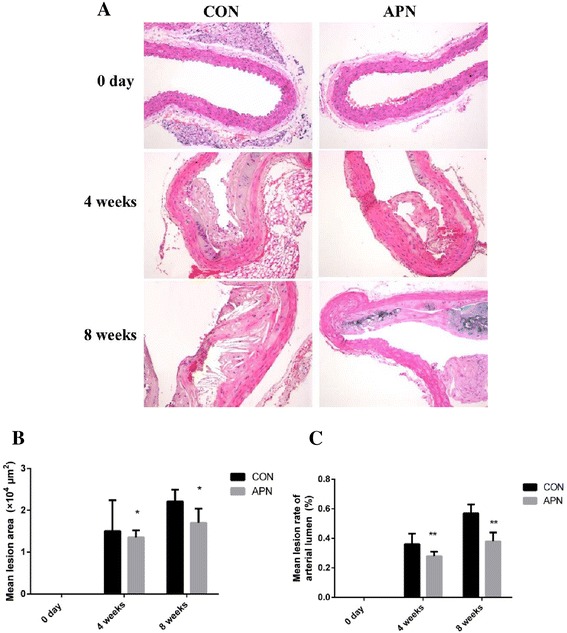
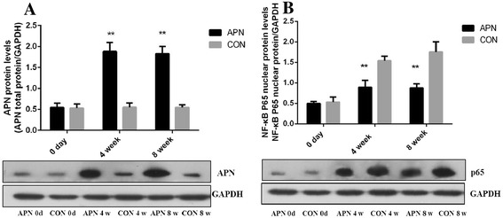
Results: Transduction of Ad-APN inhibited the formation of atherosclerotic plaque in aorta when compared with control group. The lesion formation in aortic arch root was inhibited significantly (P < 0.01). Lesion lumen ratio decreased significantly (P < 0.001). The expression of adiponectin attenuated the increases of serum TC (P < 0.001), TG (P < 0.001), and LDL-C (P < 0.001) induced by the high-fat diet, and the increase in body weight (P < 0.05). As increasing serum adiponectin, the levels of MMP-9 were significantly decreased (P < 0.05). The exogenous adiponectin increased the gene expression of the anti-inflammatory factors eNOS (P < 0.05) and IL-10 (P < 0.001), and reduced the gene expression of inflammatory factors tumor necrosis factor-α (TNF-α) (P < 0.001), IL-6 (P < 0.001), VCAM-1 (P < 0.05), respectively. Adiponectin effectively inhibited the activation of NF-κB pathway and the expression of NF-κB nuclear protein p65.

Conclusions: Adiponectin may protect the aorta from atherosclerotic injury by reducing inflammation. The molecular mechanism may involve inhibited the expression of downstream components of NF-κB and its transcription factors.

Keywords: Adiponectin; Atherosclerosis; Inflammation; NF-κB signaling pathways.


Figures

Fig. 1
Fig. 1
Overexpression of adiponectin inhibits atherosclerosis in ApoE-/- mice a Schema of experimental procedure b Lesion areas shown were quantified using Oil-red O staining of the thoracoabdominal aorta c The overexpression of adiponectin of the aortic arch root by immunohistochemical approach (×200) d Surface of lesion in thoracoabdominal aorta (%) e the number of APN+ cells/% of total (intimal/medial) area. Notes: compared with CON group, *P < 0.01, **P < 0.001, n = 6, mean ± SD
Fig. 2
Fig. 2
Mean lesion area and mean lesion rate of the aortic arch root a HE staining × 200 b The average lesion area of each mouse was determined from aortic root c The mean lesion rate. Notes: compared with CON group, *P < 0.01; **P < 0.001, n = 6, mean ± SD
Fig. 3
Fig. 3
Composition of cells in the aortic arch root a Masson trichrome staining of aortic root (×200) b Collagen content in advanced atherosclerotic plaques c Fibrous cap thickness. Notes: compared with CON group, *P < 0.01; **P < 0.001, n = 6, mean ± SD
Fig. 4
Fig. 4
Composition of cells in the aortic arch root a The area of atherosclerotic plaques in sections of the proximal aorta was stained with an antibody Mac3 against macrophages (×200) b The area of atherosclerotic plaques in sections of the proximal aorta was stained with an antibody against αSMA (×200) c the number of Mac3+ cells/% of total intimal area d the number of αSMA+ cells/% of total (intimal/medial) area. Notes: compared with CON group, #P < 0.05; **P < 0.001, n = 6, mean ± SD
Fig. 5
Fig. 5
Level of serum adiponectin、MMP-9 a Serum concentration of adiponectin. b Serum concentration of MMP-9. Notes: compared with CON group, #P < 0.05; **P < 0.001, n = 12, mean ± SD
Fig. 6
Fig. 6
a Nuclear NF-κB p65 expression in the aortic arch root in control group b Nuclear NF-κB p65 expression in the aortic arch root in adiponectin group c Expression of adiponectin、vWF in the vascular aortas in control group (×200) d Expression of adiponectin、vWF in the vascular aortas in adiponectin group (×200) e the number of APN+ cells/% of total intimal area f the number of nuclear NF-κB p65+ cells/% of total (intimal/medial) area. Notes: compared with CON group, *P < 0.01; **P < 0.001, n = 6, mean ± SD
Fig. 7
Fig. 7
a Adiponectin protein expression in the aorta b NF-κB p65 protein of nuclear expression in the aorta. Notes: compared with CON group, **P < 0.001, n = 3, mean ± SD
Fig. 8
Fig. 8
Exogenous adiponectin reduces atherosclerosis-induced inflammation in the aorta a mRNA expression of adiponectin in the vascular aortas of mice b mRNA expression of eNOS c mRNA expression of IL-10 d mRNA expression of VCAM-1 e mRNA expression of TNF-α f mRNA expression of IL-6. Notes: compared with CON group, *P < 0.01; **P < 0.001, n = 6, mean ± SD
Fig. 9
Fig. 9
Exogenous adiponectin reduces atherosclerosis-induced inflammation in the aorta a protein expression of IL-6 in the vascular aortas b protein expression of TNF-α c protein expression of VCAM-1 d protein expression of IL-10 e protein expression of eNOS. Notes: compared with CON group, *P < 0.01, n = 3, mean ± SD

References

    1. Sessa R, Pietro MD, Filardo S, Turriziani O. Infectious burden and atherosclerosis: A clinical issue. World J Clin Cases. 2014;2:240–249. doi: 10.12998/wjcc.v2.i7.240. - DOI - PMC - PubMed
    1. Viola J, Soehnlein O. Atherosclerosis - A matter of unresolved inflammation. Semin Immunol. 2015;27(3):184–93. doi: 10.1016/j.smim.2015.03.013. - DOI - PubMed
    1. Gao X, Xu XB, Belmadani S, Park Y, Tang ZH, Feldman AM, et al. TNF-alpha contributes to endothelial dysfunction by upregulating arginase in ischemia/reperfusion injury. Arterioscler Thromb Vasc Biol. 2007;27:1269–1275. doi: 10.1161/ATVBAHA.107.142521. - DOI - PubMed
    1. Takeuchi S, Wada K, Uozumi Y, Otani N, Osada H, Nagatani K, et al. Adiponectin receptor 1 expression is associated with carotid plaque stability. Neurol India. 2013;61:249–253. doi: 10.4103/0028-3886.115063. - DOI - PubMed
    1. Yamauchi T, Iwabu M, Okada-Iwabu M, Kadowaki T. Adiponectin receptors: A review of their structure, function and how they work. Best Pract Res Clin Endocrinol Metab. 2014;28:15–23. doi: 10.1016/j.beem.2013.09.003. - DOI - PubMed

Publication types

MeSH terms