. 2016 Jun;30(6):1327-34.
doi: 10.1038/leu.2016.39. Epub 2016 Feb 29.

MIM regulates the trafficking of bone marrow cells via modulating surface expression of CXCR4

Affiliations

MIM regulates the trafficking of bone marrow cells via modulating surface expression of CXCR4

T Zhan et al. Leukemia. 2016 Jun.

Abstract

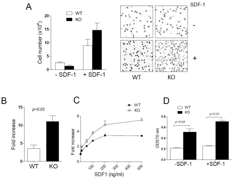
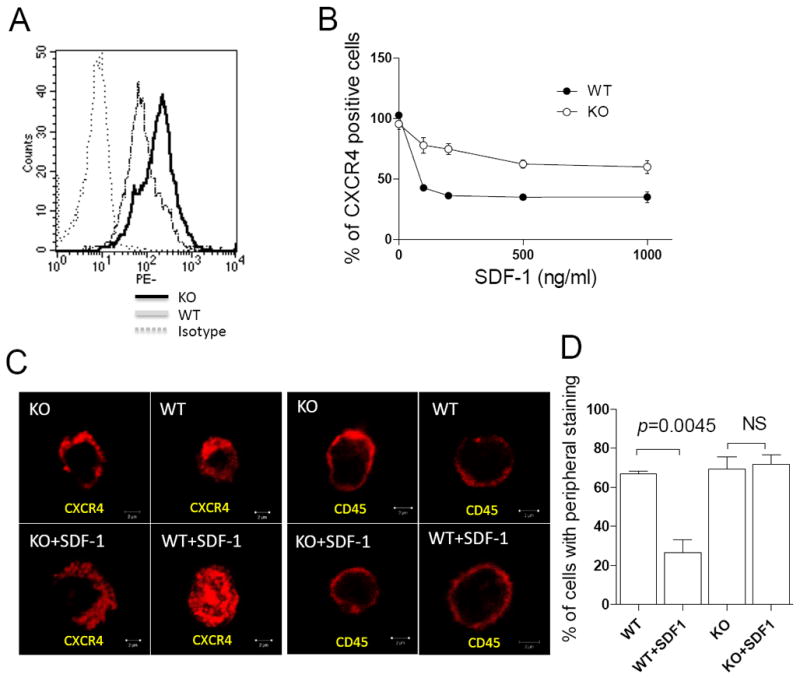
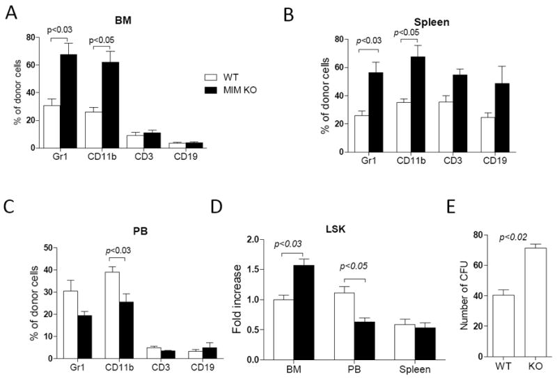
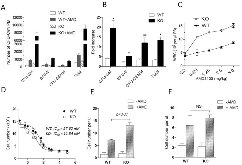
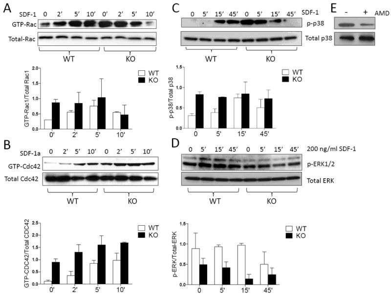
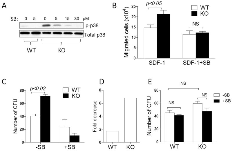
Missing in metastasis (MIM) is abundantly expressed in hematopoietic cells. Here we characterized the impact of MIM deficiency on murine bone marrow (BM) cells. Although MIM(-/-) cells proliferated similarly to wild type (WT), they exhibited stronger response to chemokine stromal-derived factor 1 (SDF-1), increase in surface expression of CXCR4, impaired CXCR4 internalization and constitutive activation of Rac, Cdc42 and p38. Transplantation of MIM(-/-) BM cells into lethally irradiated mice showed enhanced homing to BM, which was abolished when mice were pretreated with a p38 antagonist. Interestingly, MIM(-/-) BM cells, including hematopoietic stem and progenitor cells (HSPCs), showed two- to fivefold increase in mobilization into the peripheral blood upon treatment with AMD3100. In vitro, MIM(-/-) leukocytes were susceptible to AMD3100 and maintained increased response to AMD3100 for mobilization even after transfer into WT mice. MIM(-/-) mice had also a higher level of SDF-1 in the circulation. Our data highlighted an unprecedented role of MIM in the homeostasis of BM cells, including HSPCs, through modulation of the CXCR4/SDF-1 axis and interactions of BM leukocytes with their microenvironments.


Figures

Figure 1
Figure 1. MIM-/- BM cells had a higher motility in response to SDF-1 than did WT cells
(A) WT and MIM-/- (KO) BM cells were plated in the upper chamber of Transwell plates in the presence or absence of 200 ng/ml SDF-1. After 4h, cells migrated into the lower chamber were counted (left) or photographed using a 10 x objective lens (right). (B, C) The motility of cells toward either 200 ng/ml SDF-1 (B) or SDF-1 at different concentrations (C) was measured as above. The fold increase in the mobility was calculated by normalizing the number of mobilized cells to that measured in the absence of SDF-1. (D) BM cells were plated on 24-well plates pre-coated with 2 μg/ml fibronectin and incubated for 20 min. Attached cells were stained with crystal violet and quantified based on absorption at OD570nm. All the data represent mean ± SEM (n=3).
Figure 2
Figure 2. MIM-/- cells had increased in expression of CXCR4 on the surface
(A) Freshly isolated BM cells were stained by PE-CXCR4 antibody and subsequently analyzed by flow cytometry. (B) BM cells were treated with SDF-1 for 10 min at concentrations as indicated, and then analyzed by flow cytometry for the surface expression of CXCR4. All the data represent mean ± SEM (n=3). (C) BM MNCs were stained with antibody against either CXCR4 or CD45 with or without SDF-1 treatment and examined by confocal microscopy. Bar, 2 μm. (D) Quantification of BM MNCs that showed CXCR4 staining mainly at peripheral areas.
Figure 3
Figure 3. MIM-/- BM cells increased in homing during BM transplant
2×10⁶ WT or MIM-/- BM cells from CD4.2 mice were transplanted into lethally irradiated CD45.1 WT mice. The frequency of Gr1, CD11b, CD3 and CD19 cells in the donor population were determined by flow cytometry in the BM (A), the spleen (B) and the PB (C) of the recipients 24h after transplantation. Donor Lin-Scal-1+c-Kit+ (LSK) cells in the BM, the spleen and the PB of the recipients were also measured and normalized to WT donor LSK cells in the BM (D). All the data represent mean ± SEM (n=5). (E) BM cells were isolated from recipients 24 h after transplantation and analyzed by colony-forming assay (n=3). All p values were calculated by Student’s t-test.
Figure 4
Figure 4. MIM-/- cells were hypersensitive to AMD3100
(A) WT and MIM-/- mice were s.c. injected with 5 mg/kg AMD3100. After 1 h, peripheral cells were collected and measured for the colony forming activity. The numbers of colony forming units (CFU) for different lineages (CFU-GM, BFU-E, and CFU-GEMM) were presented (n=5). (B) The data in A were presented as the fold increase of CFU by comprising AMD3100-injected mice with those without AMD3100 treatment. (C) Mice were injected with different doses of AMD3100 as indicated. After 1h, total white blood cells (WBC) in the circulation of the injected mice were collected and estimated by Hemavet hematology analyzer (n=3). (D) MNCs derived from the BM of WT and MIM-/- mice were analyzed for the chemotactic response to 200 ng/ml SDF-1 in the presence of AMD3100 at the indicated concentrations. The IC50 values were calculated using Prism 5 software. (E) WT CD45.1 mice were transplanted with MNCs derived from the BM of CD45.2 WT or MIM-/- mice. The transplanted mice were then injected with 5mg/Kg AMD3100. After 1h, the amount of CD45.2+CD19+ cells in the PB of treated mice was estimated as described in the Method. The data represents mean ± SEM (n=3). (F) MNCs isolated from WT CD45.1 mice were transplanted into CD45.2 WT or MIM-/- mice (n=3). The transplanted mice were then treated with AMD3100, and the mobilized CD45.1+CD19+ cells in the PB were analyzed as above. *, p<0.002; **; p < 0.02 (t-test), referring to the difference between KO and WT mice.
Figure 5
Figure 5. MIM-/- BM cells have enhanced CXCR4 signaling
BM cells derived from WT and MIM-/- mice were treated with 200 ng/ml SDF-1 for the times as indicated and then analyzed for the presence of GTP-Rac (A), GTP-Cdc42 (B), phosphorylated p38 (C) and phosphorylated ERK1/2 (D) by Western blot. The charts below each image were the quantification results of three independent experiments. (E) Arresting BM cells of MIM-/- mice were treated with and without AMD3100 for 1h. The phosphorylated p38 was analyzed by Western blot.
Figure 6
Figure 6. p38 antagonist inhibited the increased mobility and the homing activity of MIM-/- cells
(A) MIM-/- and WT BM cells were treated for 2h with SB203580 at the concentrations as indicated and then analyzed for the level of phosphorylated p38 by Western blot. (B) WT and MIM-/- BM cells were treated with 5 μM SB203580 for 1h and analyzed for the motility response to SDF-1. The data represent mean ± SEM (n=3). (C) WT and MIM-/- BM cells were treated with 5 μM SB203580 for 1h and subsequently transplanted into lethally irradiated mice. After 24h, donor cells were isolated from the BM of recipients and analyzed for the clonogenic activity (n=2). The number of colonies was also compared between treated and non-treated cells and presented as fold decreases (D). (E) BM cells derived from WT and MIM-/- mice were treated with or without 5 μM SB203580 for 1h and then analyzed for the clonogenic activity. The data represents mean ± SEM (n=3). All the p values were based on t-test. NS, no significance.

References

    1. Ma Q, Jones D, Springer TA. The chemokine receptor CXCR4 is required for the retention of B lineage and granulocytic precursors within the bone marrow microenvironment. Immunity. 1999 Apr;10(4):463–471. - PubMed
    1. Zou YR, Kottmann AH, Kuroda M, Taniuchi I, Littman DR. Function of the chemokine receptor CXCR4 in haematopoiesis and in cerebellar development. Nature. 1998 Jun 11;393(6685):595–599. - PubMed
    1. Sugiyama T, Kohara H, Noda M, Nagasawa T. Maintenance of the hematopoietic stem cell pool by CXCL12-CXCR4 chemokine signaling in bone marrow stromal cell niches. Immunity. 2006 Dec;25(6):977–988. - PubMed
    1. Mazo IB, Massberg S, von Andrian UH. Hematopoietic stem and progenitor cell trafficking. Trends Immunol. 2011 Oct;32(10):493–503. - PMC - PubMed
    1. Burger M, Glodek A, Hartmann T, Schmitt-Graff A, Silberstein LE, Fujii N, et al. Functional expression of CXCR4 (CD184) on small-cell lung cancer cells mediates migration, integrin activation, and adhesion to stromal cells. Oncogene. 2003 Nov 6;22(50):8093–8101. - PubMed