

Identification of Susceptibility Loci and Genes for Colorectal Cancer Risk
- PMID: 26965516
- PMCID: PMC4909543
- DOI: 10.1053/j.gastro.2016.02.076
Identification of Susceptibility Loci and Genes for Colorectal Cancer Risk
Abstract
Background & aims: Known genetic factors explain only a small fraction of genetic variation in colorectal cancer (CRC). We conducted a genome-wide association study to identify risk loci for CRC.
Methods: This discovery stage included 8027 cases and 22,577 controls of East-Asian ancestry. Promising variants were evaluated in studies including as many as 11,044 cases and 12,047 controls. Tumor-adjacent normal tissues from 188 patients were analyzed to evaluate correlations of risk variants with expression levels of nearby genes. Potential functionality of risk variants were evaluated using public genomic and epigenomic databases.
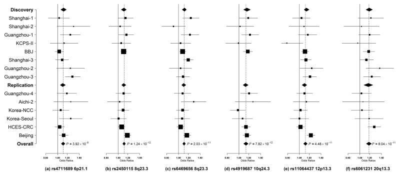
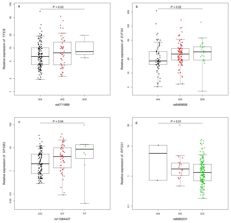
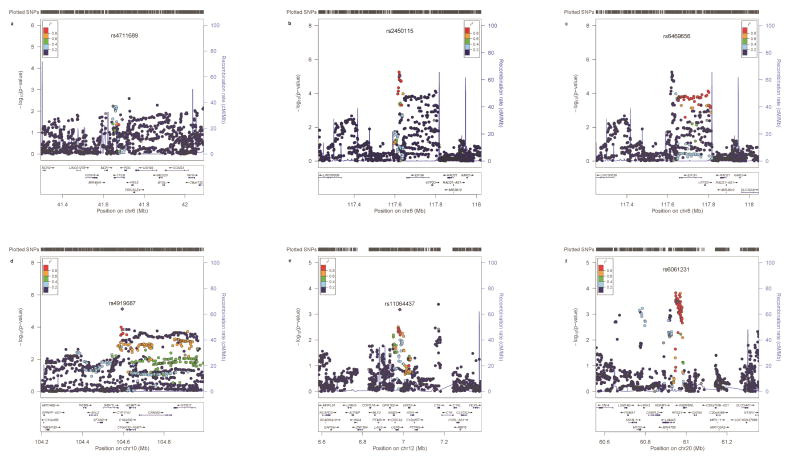
Results: We identified 4 loci associated with CRC risk; P values for the most significant variant in each locus ranged from 3.92 × 10(-8) to 1.24 × 10(-12): 6p21.1 (rs4711689), 8q23.3 (rs2450115, rs6469656), 10q24.3 (rs4919687), and 12p13.3 (rs11064437). We also identified 2 risk variants at loci previously associated with CRC: 10q25.2 (rs10506868) and 20q13.3 (rs6061231). These risk variants, conferring an approximate 10%-18% increase in risk per allele, are located either inside or near protein-coding genes that include transcription factor EB (lysosome biogenesis and autophagy), eukaryotic translation initiation factor 3, subunit H (initiation of translation), cytochrome P450, family 17, subfamily A, polypeptide 1 (steroidogenesis), splA/ryanodine receptor domain and SOCS box containing 2 (proteasome degradation), and ribosomal protein S2 (ribosome biogenesis). Gene expression analyses showed a significant association (P < .05) for rs4711689 with transcription factor EB, rs6469656 with eukaryotic translation initiation factor 3, subunit H, rs11064437 with splA/ryanodine receptor domain and SOCS box containing 2, and rs6061231 with ribosomal protein S2.
Conclusions: We identified susceptibility loci and genes associated with CRC risk, linking CRC predisposition to steroid hormone, protein synthesis and degradation, and autophagy pathways and providing added insight into the mechanism of CRC pathogenesis.
Keywords: Colon Cancer; Epidemiology; Single Nucleotide Polymorphisms; eQTL.
Copyright © 2016 AGA Institute. Published by Elsevier Inc. All rights reserved.
Figures
Comment in
-
The Hunting of the Snark: Whither Genome-Wide Association Studies for Colorectal Cancer?Gastroenterology. 2016 Jun;150(7):1528-1530. doi: 10.1053/j.gastro.2016.04.021. Epub 2016 Apr 29. Gastroenterology. 2016. PMID: 27133396 No abstract available.
References
-
- Torre LA, Bray F, Siegel RL, et al. Global cancer statistics, 2012. CA Cancer J Clin. 2015;65:87–108. - PubMed
-
- Lichtenstein P, Holm NV, Verkasalo PK, et al. Environmental and heritable factors in the causation of cancer - Analyses of cohorts of twins from Sweden, Denmark, and Finland. New England Journal of Medicine. 2000;343:78–85. - PubMed
MeSH terms
Substances
Grants and funding
- U01 HG004438/HG/NHGRI NIH HHS/United States
- U01 HG004446/HG/NHGRI NIH HHS/United States
- K05 CA154337/CA/NCI NIH HHS/United States
- HHSN268201100046C/HL/NHLBI NIH HHS/United States
- R01 CA064277/CA/NCI NIH HHS/United States
- UM1 CA186107/CA/NCI NIH HHS/United States
- HHSN268201100002C/WH/WHI NIH HHS/United States
- R01 CA042182/CA/NCI NIH HHS/United States
- UM1 CA167552/CA/NCI NIH HHS/United States
- HHSN268201100003I/HL/NHLBI NIH HHS/United States
- HHSN268201100004C/WH/WHI NIH HHS/United States
- U19 CA148107/CA/NCI NIH HHS/United States
- R01 CA059045/CA/NCI NIH HHS/United States
- HHSN268201100001I/HL/NHLBI NIH HHS/United States
- R01 CA076366/CA/NCI NIH HHS/United States
- R01 CA188214/CA/NCI NIH HHS/United States
- U01 CA074799/CA/NCI NIH HHS/United States
- P50 CA095103/CA/NCI NIH HHS/United States
- R01 CA082729/CA/NCI NIH HHS/United States
- P01 CA087969/CA/NCI NIH HHS/United States
- HHSN268201100004I/HL/NHLBI NIH HHS/United States
- Z01 CP010200/ImNIH/Intramural NIH HHS/United States
- P01 CA055075/CA/NCI NIH HHS/United States
- R37 CA070867/CA/NCI NIH HHS/United States
- R01 CA151993/CA/NCI NIH HHS/United States
- R01 CA048998/CA/NCI NIH HHS/United States
- U01 CA137088/CA/NCI NIH HHS/United States
- HHSN268201100003C/WH/WHI NIH HHS/United States
- T32 HG000040/HG/NHGRI NIH HHS/United States
- UM1 CA173640/CA/NCI NIH HHS/United States
- U01 CA164930/CA/NCI NIH HHS/United States
- R01 CA092585/CA/NCI NIH HHS/United States
- R01 CA137178/CA/NCI NIH HHS/United States
- U01 CA074794/CA/NCI NIH HHS/United States
- P30 CA014089/CA/NCI NIH HHS/United States
- R01 CA148667/CA/NCI NIH HHS/United States
- R01 CA081488/CA/NCI NIH HHS/United States
- HHSN271201100004C/AG/NIA NIH HHS/United States
- R01 CA124558/CA/NCI NIH HHS/United States
- P30 CA068485/CA/NCI NIH HHS/United States
- U01 CA097735/CA/NCI NIH HHS/United States
- T32 ES013678/ES/NIEHS NIH HHS/United States
- UM1 CA167551/CA/NCI NIH HHS/United States
- U01 CA122839/CA/NCI NIH HHS/United States
- HHSN268201100002I/HL/NHLBI NIH HHS/United States
- U01 CA074783/CA/NCI NIH HHS/United States
- U01 CA074806/CA/NCI NIH HHS/United States
- UM1 CA182910/CA/NCI NIH HHS/United States
- P50 CA127003/CA/NCI NIH HHS/United States
- P30 CA071789/CA/NCI NIH HHS/United States
- U01 CA074800/CA/NCI NIH HHS/United States
- HHSN268201100001C/WH/WHI NIH HHS/United States
LinkOut - more resources
Full Text Sources
Other Literature Sources
Medical
