. 2016 Aug;100(2):391-401.
doi: 10.1189/jlb.3A1015-480R. Epub 2016 Mar 10.

CD63 is tightly associated with intracellular, secretory events chaperoning piecemeal degranulation and compound exocytosis in human eosinophils

Affiliations

CD63 is tightly associated with intracellular, secretory events chaperoning piecemeal degranulation and compound exocytosis in human eosinophils

Lívia A S Carmo et al. J Leukoc Biol. 2016 Aug.

Abstract

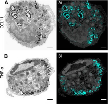
Eosinophil activation leads to secretion of presynthesized, granule-stored mediators that determine the course of allergic, inflammatory, and immunoregulatory responses. CD63, a member of the transmembrane-4 glycoprotein superfamily (tetraspanins) and present on the limiting membranes of eosinophil-specific (secretory) granules, is considered a potential surface marker for eosinophil degranulation. However, the intracellular secretory trafficking of CD63 in eosinophils and other leukocytes is not understood. Here, we provide a comprehensive investigation of CD63 trafficking at high resolution within human eosinophils stimulated with inflammatory stimuli, CCL11 and tumor necrosis factor α, which induce distinctly differing secretory processes in eosinophils: piecemeal degranulation and compound exocytosis, respectively. By using different transmission electron microscopy approaches, including an immunonanogold technique, for enhanced detection of CD63 at subcellular compartments, we identified a major intracellular pool of CD63 that is directly linked to eosinophil degranulation events. Transmission electron microscopy quantitative analyses demonstrated that, in response to stimulation, CD63 is concentrated within granules undergoing secretion by piecemeal degranulation or compound exocytosis and that CD63 tracks with the movements of vesicles and granules in the cytoplasm. Although CD63 was observed at the cell surface after stimulation, immunonanogold electron microscopy revealed that a strong CD63 pool remains in the cytoplasm. It is remarkable that CCL11 and tumor necrosis factor α triggered increased formation of CD63(+) large vesiculotubular carriers (eosinophil sombrero vesicles), which fused with granules in the process of secretion, likely acting in the intracellular translocation of CD63. Altogether, we identified active, intracellular CD63 trafficking connected to eosinophil granule-derived secretory pathways. This is important for understanding the complex secretory activities of eosinophils underlying immune responses.

Keywords: cell secretion; immune responses; inflammation; transmission electron microscopy; vesicular trafficking.


Figures

Figure 1
Figure 1
CD63 Immunolabeling in nonpermeabilized human eosinophils. Although unstimulated cells (A) show absent or weak fluorescence, a pool of CD63 imaged as green fluorescence is observed at the surface of cells stimulated with CCL11 (B) or TNF‐α (C). Note that the immunoreactivity pattern is punctate with CCL11 (B) and mostly diffuse with TNF‐α (C). Eosinophils were isolated by negative selection from the blood of healthy human donors, stimulated with CCL11 (B) or TNF‐α (C) for 1 h, and incubated with mouse anti‐human CD63 or isotype antibody, followed by secondary antibody (anti‐mouse conjugated with Alexa Fluor 488). Control cells were kept in medium (A). Images are representative of 3 independent experiments. Scale bar, 2.3 μm (A–C).
Figure 2
Figure 2
Conventional TEM identifies distinct, secretory processes, triggered by CCL11 and TNF‐α stimulation. (A) PMD, characterized by progressive emptying of the secretory granules in the absence of granule fusions, was observed in response to CCL11. In (Ai–Aiii), note, in high magnification, the disarrangement of the granule cores and matrices. EoSVs, highlighted in pink, are seen around emptying granules. (B and C) Compound exocytosis, characterized by large channels formed by granule–granule fusions, was the predominant mode of secretion induced by TNF‐α. In (D), granule losses and fusion of the first granule from the channel with the plasma membrane (arrow) are shown. (E) Significant increases in numbers of emptying or fused granules occurred after stimulation with CCL11 or TNF‐α, respectively. Eosinophils were isolated from the peripheral blood by negative selection, immediately fixed, and processed for conventional TEM. Counts were derived from 3 experiments with 3259 granules counted in 87 electron micrographs randomly taken and showing the entire cell profile and nucleus. Scale bar, 700 nm (A), 315 nm (Ai–Aiii), 860 nm (B), 1.1 μm (C), and 580 nm (D). Data represent means ± sem ****P < 0.0001 vs. control intact granules; #### P < 0.0001 vs. control‐emptying granules; ++++ P < 0.0001 vs. control‐fused granules.
Figure 3
Figure 3
CD63 immunolabeling of secretory granules by immunonanogold EM. (A) Quantitative EM analyses revealed that most secretory granules were positive for CD63 in all groups. Stimulation led to significant increase in the numbers of CD63‐labeled granules compared to unstimulated cells. (B) A representative electron micrograph of an unstimulated human eosinophil revealed CD63 labeling on the granules (Gr) limiting membranes. The boxed area in (B) is shown in higher magnification in (Bi). A CD63+ EoSV is indicated (arrow). Eosinophils were isolated from the peripheral blood, stimulated or not with CCL11 or TNF‐α and prepared for pre‐embedding immunanogold EM. Counts were derived from, at least, 3 experiments with 2005 granules counted in 54 electron micrographs randomly taken and showing the entire cell profile and nucleus (N). Scale bars, 800 nm (B); 500 nm (Bi). ****P < 0.0001 vs. control cells.
Figure 4
Figure 4
CD63 is strongly associated with secretory processes within human eosinophils. (A, B) Secretory granules (Gr) undergoing content release through PMD (A) or compound exocytosis (B) were intensely labeled for CD63. Note that although CD63+ granules were distributed in the entire cytoplasm in PMD (A), these organelles were concentrated in the peripheral cytoplasm in compound exocytosis (B). In (Ai and Bi), granules were highlighted in blue using Photoshop software. Cells were isolated from the peripheral blood, stimulated with CCL11 (A) or TNF‐α (B) and prepared for pre‐embedding immunanogold EM. Scale bars, 915 nm (A, Ai), 680 nm (B, Bi).
Figure 5
Figure 5
Differential distribution of CD63+ secretory granules within human eosinophils after stimulation with TNF‐α. (A) A representative electron micrograph shows several cell sections with cytoplasmic CD63+ granules, mostly confined at the cell periphery, near the plasma membrane. Arrowheads indicate CD63 polarization on the granule face toward the plasma membrane. (B) Quantitative analyses of immunolabeled granules. Eosinophils were isolated from the peripheral blood, stimulated with TNF‐α, and prepared for pre‐embedding immunonanogold EM. A total of 460 granules from 10 sharp, cross‐cell sections exhibiting the entire cell profile, intact plasma membrane, and nucleus were counted in the peripheral cytoplasm (1.0 μm wide from the plasma membrane, as indicated by the red bars) and within the inner cytoplasm (the contiguous cytoplasmic area deeper in the cell). Scale bar, 1.3 μm. ****P < 0.0001 vs. CD63+ granules at cell inner area; #### P < 0.0001 vs. CD63 granules at cell inner area.
Figure 6
Figure 6
CD63 concentrates within granules undergoing active processes of secretion. (A) Representative CD63 expression in non‐stimulated (NS), CCL11‐ or TNF‐α‐stimulated human eosinophils evaluated by western blotting (n = 3). L = lane. (B) Representative images of secretory granules at high resolution within human eosinophils after stimulation or not. Note that CD63 was concentrated within stimulated granules while in NS granules (intact) the labeling was mostly observed at the granule limiting membrane. (C) The total granule area as well as the CD63‐immunolabeled area increased in response to stimulation (*; #P < 0.05 vs. NS). In (D), the variation of CD63‐immunolabeled area in specific granules is shown in different groups. Eosinophils were isolated from the peripheral blood, stimulated or not with CCL11 or TNF‐α and prepared for pre‐embedding immunanogold EM. A total of 175 secretory (specific) granules showing pools of CD63 from CCL11‐stimulated or TNF‐α–stimulated cells and controls (n = 29 cells) were analyzed for area quantification. Scale bar, 600 nm. Data represent means ± sem.
Figure 7
Figure 7
Vesicular trafficking of CD63 within human, stimulated eosinophils. (A and B) EoSVs (highlighted in pink) were observed in the cytoplasm surrounding or fused with secretory granules (Gr) within CCL11‐stimulated (A) or TNF‐α–stimulated cells. (C) Quantitative analyses of EoSV numbers. Note that, after stimulation, not only did the total number of EoSVs increase but also the number of CD63+ EoSVs. (D) Many EoSVs were seen contacting granules undergoing secretion. (E) After stimulation with TNF‐α, most CD63+ EoSVs were observed in the cell periphery. Eosinophils were isolated from the peripheral blood, stimulated or not with CCL11 or TNF‐α and prepared for pre‐embedding immunanogold EM. A total of 23 electron micrographs from unstimulated and stimulated cells were evaluated, and the numbers of labeled and not labeled EoSVs (n = 1945) were counted in each cell section. NS, not stimulated. *P < 0.05 vs. NS group (total EoSVs number); # P < 0.05 vs. NS group (CD63+ EoSVs); ##P < 0.01 vs. NS group (CD63+ EoSVs); ***P < 0.001 vs. CD63+ EoSVs at cell inner. Scale bar, 437 nm (A and Ai). Data represent means ± sem.
Figure 8
Figure 8
CD63 is translocated on EoSVs to or from secretory granules after stimulation with TNF‐α. (A and Ai) A representative electron micrograph from an entire eosinophil profile shows CD63‐labeled EoSVs (highlighted in pink), mostly at cell periphery, in association with CD63+ secretory granules (Gr). The boxed area in (A) is shown in higher magnification in (Ai). Eosinophils were isolated from peripheral blood, stimulated with TNF‐α, and prepared for pre‐embedding immunanogold EM. N, nucleus. Scale bar, 950 nm (A), 630 nm (Ai).

Similar articles

Cited by

References

    1. Stow, J. L. , Murray, R. Z. (2013) Intracellular trafficking and secretion of inflammatory cytokines. Cytokine Growth Factor Rev. 24, 227–239. - PubMed
    1. Spencer, L. A. , Bonjour, K. , Melo, R. C. , Weller, P. F. (2014) Eosinophil secretion of granule‐derived cytokines. Front. Immunol. 5, 496. - PMC - PubMed
    1. Melo, R. C. N. , Dvorak, A. M. , Weller, P. F. 2012. Eosinophil ultrastructure In Eosinophils in health and disease. (Lee J. R., Rosenberg H., eds.), Vol. 1 Elsevier, New York, p. 20–27.
    1. McLaren, D. J. , Mackenzie, C. D. , Ramalho‐Pinto, F. J. (1977) Ultrastructural observations on the in vitro interaction between rat eosinophils and some parasitic helminths (Schistosoma mansoni, Trichinella spiralis and Nippostrongylus brasiliensis). Clin. Exp. Immunol. 30, 105–118. - PMC - PubMed
    1. Inoue, Y. , Matsuwaki, Y. , Shin, S. H. , Ponikau, J. U. , Kita, H. (2005) Nonpathogenic, environmental fungi induce activation and degranulation of human eosinophils. J. Immunol. 175, 5439–5447. - PubMed

Publication types