. 2016 Mar 11:6:22966.
doi: 10.1038/srep22966.

The Wnt/β-catenin signaling/Id2 cascade mediates the effects of hypoxia on the hierarchy of colorectal-cancer stem cells

Affiliations

The Wnt/β-catenin signaling/Id2 cascade mediates the effects of hypoxia on the hierarchy of colorectal-cancer stem cells

Hye-Jin Dong et al. Sci Rep. .

Abstract

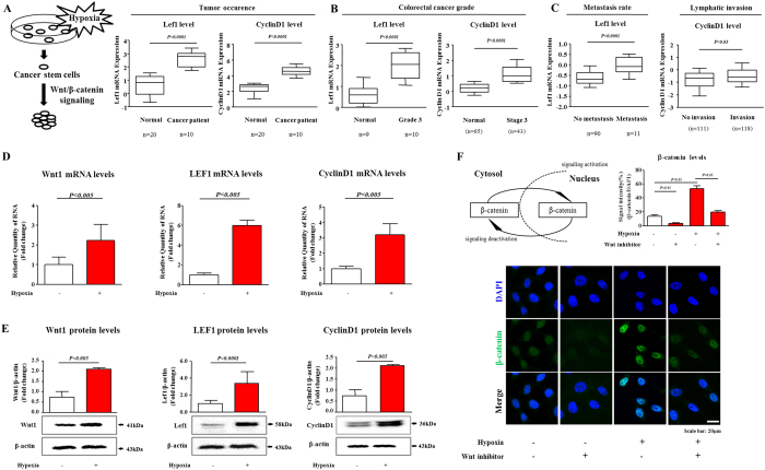
Hypoxia, a feature common to most solid tumors, is known to regulate many aspects of tumorigenesis. Recently, it was suggested that hypoxia increased the size of the cancer stem-cell (CSC) subpopulations and promoted the acquisition of a CSC-like phenotype. However, candidate hypoxia-regulated mediators specifically relevant to the stemness-related functions of colorectal CSCs have not been examined in detail. In the present study, we showed that hypoxia specifically promoted the self-renewal potential of CSCs. Through various in vitro studies, we found that hypoxia-induced Wnt/β-catenin signaling increased the occurrence of CSC-like phenotypes and the level of Id2 expression in colorectal-cancer cells. Importantly, the levels of hypoxia-induced CSC-sphere formation and Id2 expression were successfully attenuated by treatment with a Wnt/β-catenin-signaling inhibitor. We further demonstrated, for the first time, that the degree of hypoxia-induced CSC-sphere formation (CD44(+) subpopulation) in vitro and of tumor metastasis/dissemination in vivo were markedly suppressed by knocking down Id2 expression. Taken together, these data suggested that Wnt/β-catenin signaling mediated the hypoxia-induced self-renewal potential of colorectal-cancer CSCs through reactivating Id2 expression.


Figures

Figure 1
Figure 1. The effects of hypoxia on Wnt/β-catenin signaling in colorectal-cancer cells.
(A–C) A significant correlation between tumor development/metastasis and the expression of Wnt/β-catenin-signaling components was observed in the human colorectal-cancer datasets available through the Oncomine dataset repository (www.oncomine.org). Real-time PCR (D) and western blotting (E) demonstrated the hypoxia-induced changes in the expression of Wnt/β-catenin-signaling components (Wnt1, Lef1, and cyclin D1). (F) Colorectal-cancer cells were stained using an antibody specific for β-catenin. The stimulatory effects of hypoxia on these activities of β-catenin were successfully attenuated after Wnt/β-catenin-signaling inhibitor treatment. DAPI staining was used to label the nuclei. β-actin was used as the internal control. The results are the mean values ± SD from three independent experiments.
Figure 2
Figure 2. Stimulatory effect of hypoxia on the growth and the stemness-related characteristics of colorectal CSCs.
(A) Hypoxia stimulated CSC-sphere formation by the mouse colorectal-cancer cells after 4 days under sphere-formation culturing conditions. The number of spheres that were larger than 150 μm in diameter was enumerated, and a representative image of the CSC-containing spheres is shown. The data represents the average values from three independent experiments. (B) Real-time PCR data demonstrated the hypoxia-induced enhanced expression of the stem-cell markers c-Myc and Sox2. (C) The results of FACS analysis showed the increased percentage of CD44-positive cells among the total population of mouse colorectal-cancer cells grown under the hypoxic condition. (D) The stimulatory effects of hypoxia on colorectal CSC-sphere formation were successfully attenuated by treatment with a Wnt/β-catenin-signaling inhibitor. (E) Treatment with a Wnt/β-catenin-signaling inhibitor led to the decrease in the percentage of CD44-positive cells in the colorectal-cancer cells grown under hypoxic conditions. The results are the mean values ± SD from three independent experiments.
Figure 3
Figure 3. Knocking down Id2 expression suppressed the hypoxia-induced increased growth and exhibition of stemness-related characteristics of colorectal CSCs.
(A) Mouse colorectal-cancer cells cultured under the hypoxic condition were evaluated for the expression level of Id family members (Id1-Id4). (B) The stimulatory effect of hypoxia on the expression of Id2 was verified by western blotting analysis. (C) The results of real-time PCR and (D) western blotting assays demonstrated the change of Id2 expression that significantly increased by Wnt-ligand treatment. The levels of expression of Id2 in mouse colorectal-cancer cells treated with the Wnt/β-catenin-signaling inhibitor (2 μM) with or without hypoxia were evaluated using (E) real-time PCR and (F) western blotting. (G) Knocking down Id2 expression successfully attenuated the hypoxia-induced CSC-sphere formation. The number of spheres that were larger than 150 μm in diameter was counted, and a representative image of a tumorsphere is shown. The Id2 knockdown was verified based on the (H) mRNA and (I) protein levels observed under our colorectal CSC-culturing conditions. β-actin was used as the internal control. Abbreviations: TSFE, tumor sphere-forming efficiency. The results are the mean values ± SD from three independent experiments.
Figure 4
Figure 4. The effects of the Id2 knockdown on colorectal cancer-cell growth and apoptosis.
(A–C) A significant correlation between tumor development/metastasis and Id2 expression were observed in human colorectal-cancer datasets available through the Oncomine dataset repository (www.oncomine.org). (D) Transfection of colorectal-cancer cells with Id2 shRNA led to a time-dependent decrease in the number of cells compared with transfection with the control shRNA. (E) Id2 knockdown-mediated apoptotic DNA fragmentation and condensation were visualized using DAPI staining. (F) The Id2 knockdown-mediated cytotoxicity was evaluated by flow cytometry using PE-labeled anti-annexin-V. (G) The level of activated (cleaved) caspase 3 in the cells undergoing Id2 knockdown-induced apoptotic death was evaluated by western blotting using an antibody directed against activated caspase 3. DAPI staining was used to label the nuclei. β-actin was used as the internal control. The results are the mean values ± SD from three independent experiments.
Figure 5
Figure 5. The effects of the Id2 knockdown on the invasion and migration of colorectal-cancer cells.
(A) The cell-invasion ability of the colorectal-cancer cells was evaluated using a transwell assay. Transfection with Id2 shRNA significantly decreased the degree of their invasion across the transwell membrane compared with transfection with the control shRNA. (B) The effects of the Id2 knockdown on the migration of colorectal-cancer cells were evaluated using a scratch assay. The migration of cells transfected with the shRNA targeting Id2 was slower than that of the cells transfected with the control nontargeting shRNA. (C,D) The relative level of expression of the migration regulators MMP2 and MMP9 was assessed using real-time PCR and western blot. (E) The Id2 knockdown-induced disorganization of actin filaments and the morphological transition of the cells were visualized through phalloidin staining of actin filaments. DAPI staining was used to label the nuclei. β-actin was used as the internal control. The results are the mean values ± SD from three independent experiments.
Figure 6
Figure 6
The effects of knocking down Id2 expression in colorectal-cancer cells on their metastasis/dissemination (A,B) Mouse colorectal-cancer cells expressing firefly luciferase (CT26-Luc) were subcutaneously injected into BALB/c mice. After their injection, the abdominal metastasis/dissemination was monitored using an IVIS; the photon intensity, a measurement of the dissemination of viable tumor cells, was also determined by the system. The results are the mean values ± SD from three independent experiments.
Figure 7
Figure 7. Schematic summary of the role of the Wnt/β-catenin signaling/Id2 cascade in the development of colorectal cancer under hypoxia.
The hypoxia-induced enhancement of the Wnt/β-catenin signaling/Id2 cascade up-regulates the self-renewal and migration abilities of colorectal CSCs, thereby promoting the growth and dissemination of colorectal cancer cells.

References

    1. Vaupel P. Hypoxia and aggressive tumor phenotype: implications for therapy and prognosis. Oncologist 13 Suppl 3, 21–6 (2008). - PubMed
    1. Visvader J. E. & Lindeman G. J. Cancer stem cells: current status and evolving complexities. Cell Stem Cell 10, 717–28 (2012). - PubMed
    1. Li X. et al. Intrinsic resistance of tumorigenic breast cancer cells to chemotherapy. J Natl Cancer Inst 100, 672–9 (2008). - PubMed
    1. Bao S. et al. Glioma stem cells promote radioresistance by preferential activation of the DNA damage response. Nature 444, 756–60 (2006). - PubMed
    1. Heddleston J. M. et al. Hypoxia inducible factors in cancer stem cells. Br J Cancer 102, 789–95 (2010). - PMC - PubMed

Publication types

MeSH terms