. 2016 Mar 11:6:22971.
doi: 10.1038/srep22971.

Vaginal Fibroblastic Cells from Women with Pelvic Organ Prolapse Produce Matrices with Increased Stiffness and Collagen Content

Affiliations

Vaginal Fibroblastic Cells from Women with Pelvic Organ Prolapse Produce Matrices with Increased Stiffness and Collagen Content

Alejandra M Ruiz-Zapata et al. Sci Rep. .

Abstract

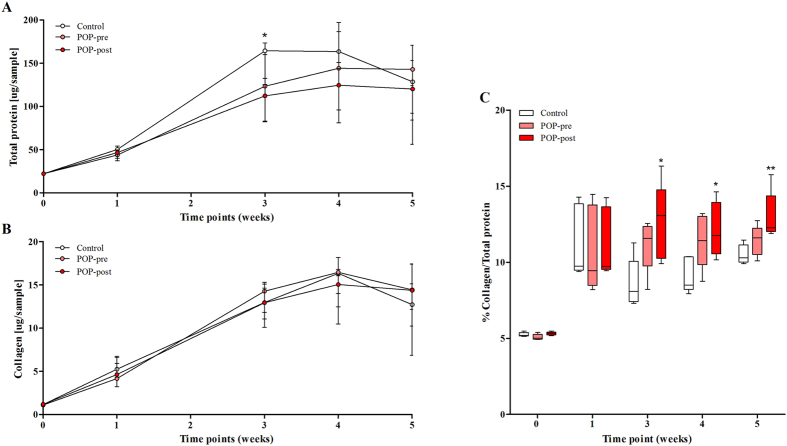
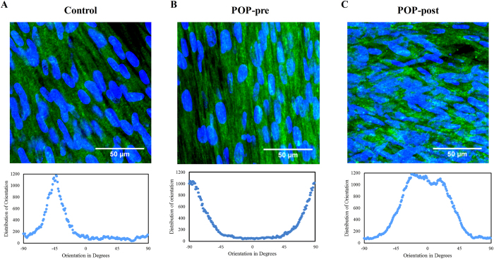
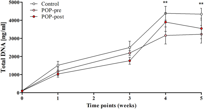
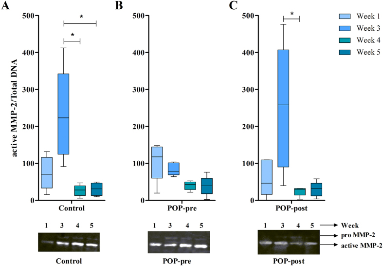
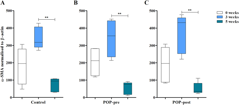
Pelvic organ prolapse (POP) is characterised by the weakening of the pelvic floor support tissues, and often by subsequent prolapse of the bladder outside the body, i.e. cystocele. The bladder is kept in place by the anterior vaginal wall which consists of a dense extracellular matrix rich in collagen content that is maintained and remodelled by fibroblastic cells, i.e. fibroblasts and myofibroblasts. Since altered matrix production influences tissue quality, and myofibroblasts are involved in normal and pathological soft tissue repair processes, we evaluated matrix production of cells derived from pre- and post-menopausal POP and non-POP control anterior vaginal wall tissues. Results showed that cells from postmenopausal POP women deposited matrices with high percentage of collagen fibres with less anisotropic orientation and increased stiffness than those produced by controls. There was a transient increase in myofibroblastic phenotype that was lost after the peak of tissue remodelling. In conclusion, affected fibroblasts from postmenopausal prolapsed tissues produced altered matrices in vitro compared to controls. Such aberrant altered matrix production does not appear to be a consequence of abnormal phenotypical changes towards the myofibroblastic lineage.


Figures

Figure 1
Figure 1. Fibroblastic cells from postmenopausal prolapsed tissues deposit less extracellular matrix than controls, but with high collagen content.
Cells derived from prolapsed tissues from premenopausal (POP-pre) and postmenopausal (POP-post) women were cultured in vitro for five weeks in the presence of vitamin C and compared to controls. The deposited matrices were stained with Sirius red/fast green collagen staining kit (Chondrex Inc.) at different time points. Data represent the median (+/− interquartile range) of five samples per group and is shown as: (A) total protein (control vs. POP-post: p = 0.0486), (B) collagen content, and (C) box plots of the percentage (%) of collagen content per total protein (control vs. POP-post at week 3, p = 0.0327; at week 4, p = 0.0400; and week 5, p = 0.0089). Differences between control and POP-post were identified by Kruskal-Wallis test followed by Dunn’s multiple comparison: *p < 0.05 and **p < 0.01.
Figure 2
Figure 2. Collagen type I fibres and cell nuclei were aligned in one preferential direction in control and premenopausal POP groups but not in the postmenopausal POP group.
Representative immunohistochemistry micrographs of collagen I (green) and cells nuclei (blue) in deposited matrices after five weeks of culture of cells derived from (A) control, (B) prolapsed premenopausal (POP-pre), and (C) prolapsed postmenopausal (POP-post). The figure depicts the maximum intensity projection from z-stacks (top panel) and their corresponding fiber orientation (bottom panel). Images were acquired with 63 x objective of a Leica microscope. The bar is 50 μm.
Figure 3
Figure 3. Proliferation of anterior vaginal wall fibroblastic cells was not affected by POP or menopausal status in vitro.
The total DNA of fibroblastic cells was measured by CyQuant at different time points. The cells were derived from prolapsed tissues from premenopausal (POP-pre) and postmenopausal (POP-post) women, as well as from controls. The box plots correspond to five samples per group per time point. **p < 0.01 by Kruskal-Wallis test followed by Dunn’s multiple comparison compared to week 0. Week 0 vs. week 4: control, p = 0.0019; POP-pre, p = 0.0069; POP-post, p = 0.0011. Week 0 vs. week 5: control, p = 0.0019; POP-pre, p = 0.0031; POP-post, p = 0.0031. No differences were found between the study groups.
Figure 4
Figure 4. Vaginal fibroblastic cells secreted and activated matrix metalloproteinases-2 in long term cultures in vitro.
Conditioned media of fibroblastic cells from prolapsed premenopausal (POP-pre) (B) and postmenopausal (POP-post) (C) tissues was evaluated at different time points and compared to controls (A). Matrix metalloproteinase (MMP)-2 was detected by zymography, in samples derived from the same experiment, with gels processed in parallel. The blots were quantified by densiometry analysis of the bands and data were normalised to total DNA. The box plots correspond to five samples per group per time point. *p < 0.05 by Kruskal-Wallis test followed by Dunn’s multiple comparison. Week 3 vs. week 4: control, p = 0.0116; and POP-post, p = 0.0234. No differences were found between the study groups. Full-length gels are presented in Supplementary Figure S1.
Figure 5
Figure 5. Vaginal fibroblasts show transient differentiation to myofibroblasts in vitro.
Cell lysates were collected at different time points (0, 3 and 5 weeks) and myofibroblast differentiation was detected by western immunoblotting of α-smooth muscle actin (α-SMA) and β-actin was used as a control. Cells originated from three groups: (A) controls, (B) prolapsed premenopausal (POP-pre), and (C) prolapsed postmenopausal (POP-post). Samples derived from the same experiment were processed in gels in parallel. Blots were quantified by densiometry analysis of the bands and data was normalised to the first band of β-actin per blot. The box plots correspond to five samples per time point per group. **p < 0.01 by Kruskal-Wallis test followed by Dunn’s multiple comparison. Week 3 vs. week 4: control, p = 0.0056; POP-pre, p = 0.0066; POP-post, p = 0.0044 No differences were found between the study groups. Full-length blots are presented in Supplementary Figure S2.

References

    1. Hendrix S. L. et al. Pelvic organ prolapse in women’s health initiative: gravity and gravidity. Am. J. Obstet. Gynecol. 186, 1160–1166 (2002). - PubMed
    1. Jelovsek J. E., Maher C. & Barber M. D. Pelvic organ prolapse. Lancet 396, 1027–1038 (2007). - PubMed
    1. Lensen E. J. M., Withagen M. I. J., Kluivers K. B., Milani A. L. & Vierhout M. E. Surgical treatment of pelvic organ prolapse: a historical review with emphasis on the anterior compartment. Int. Urogynecol. J. 24, 1593–1602 (2013). - PubMed
    1. Cartwright R. et al. Systematic review and metaanalysis of genetic association studies of urinary symptoms and prolapse in women. Am. J. Obstet. Gynecol. 212, 199.e1–199.e24 (2015). - PMC - PubMed
    1. Ashton-Miller J. A. & DeLancey J. O. On the biomechanics of vaginal birth and common sequelae. Annu. Rev. Biomed. Eng. 11, 163–176 (2009). - PMC - PubMed

Publication types