. 2016 Apr 5;113(14):3809-14.
doi: 10.1073/pnas.1522674113. Epub 2016 Mar 10.

Cytochrome c folds through foldon-dependent native-like intermediates in an ordered pathway

Affiliations

Cytochrome c folds through foldon-dependent native-like intermediates in an ordered pathway

Wenbing Hu et al. Proc Natl Acad Sci U S A. .

Abstract

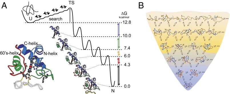
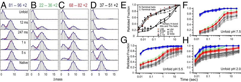
Previous hydrogen exchange (HX) studies of the spontaneous reversible unfolding of Cytochrome c (Cyt c) under native conditions have led to the following conclusions. Native Cyt c (104 residues) is composed of five cooperative folding units, called foldons. The high-energy landscape is dominated by an energy ladder of partially folded forms that differ from each other by one cooperative foldon unit. The reversible equilibrium unfolding of native Cyt c steps up through these intermediate forms to the unfolded state in an energy-ordered sequence, one foldon unit at a time. To more directly study Cyt c intermediates and pathways during normal energetically downhill kinetic folding, the present work used HX pulse labeling analyzed by a fragment separation-mass spectrometry method. The results show that 95% or more of the Cyt c population folds by stepping down through the same set of foldon-dependent pathway intermediates as in energetically uphill equilibrium unfolding. These results add to growing evidence that proteins fold through a classical pathway sequence of native-like intermediates rather than through a vast number of undefinable intermediates and pathways. The present results also emphasize the condition-dependent nature of kinetic barriers, which, with less informative experimental methods (fluorescence, etc.), are often confused with variability in intermediates and pathways.

Keywords: cytochrome c; foldons; protein folding.


Conflict of interest statement

The authors declare no conflict of interest.

Figures

Fig. 1.
Fig. 1.
Alternative folding models. (A) The defined pathway model previously inferred from HX studies of the foldon-dependent interconversion of native Cyt c with partially unfolded forms (PUFs) in the high free energy space above the native protein (10, 11, 13). (B) A representation of the energetically downhill multiple pathway model. Redrawn from ref. . These models are tested in the present work during kinetic folding.
Fig. 2.
Fig. 2.
Cooperative foldon units from unfolding experiments. (A) The native-state HX results that first defined Cyt c foldons and their cooperative unfolding. Reprinted from ref. . HX rates of many amide protons measured by 2D NMR at low denaturant concentrations are plotted in terms of the free energy of their determining exposure reactions. The ΔG of each exposure reaction, its size, and its identity can be calculated as described in the main text. (B) Illustrative HX MS results (complete data are provided in Fig. S1) that more clearly display the lower-lying red and infrared (black) foldon units by their equilibrium EX1 unfolding. (C) Time dependence for the equilibrium EX1 unfolding of many infrared and red peptides [pH 10, 10 °C, 0.22 M guanidinium chloride (GdmCl)]. (D) Illustrative stability labeling experiment used to determine the identity of the intermediate PUFs in terms of the foldons that are folded and unfolded in each state (26, 27). The unfolding ΔG measured as in A for each foldon unfolding is placed on a free energy ladder. The E62G mutation removes a salt link and destabilizes the yellow foldon unit by 1 kcal/mol (12). The green and blue foldon unfolded states are equally destabilized, indicating that they include the unfolded state of the yellow foldon. Lower-lying PUFs are unaffected, indicating that their unfolding does not include the unfolded yellow foldon. A set of similar experiments indicates that the identity of the PUFs is IRYGB (=N), iRYGB, irYGB, iryGB, irygB, and irygb (= U).
Fig. 3.
Fig. 3.
Kinetic folding of Cyt c. (AD) Illustrative mass spectra for some peptides from HX pulse labeling experiments during Cyt c folding (more complete data are in Fig. S2). Initially unfolded fully deuterated Cyt c was diluted into folding conditions at pH 5 and probed during folding by a high-pH D to H labeling pulse. The isotopic envelope for each peptide is composed of peptide molecules that carry differing numbers of 2H atoms (variable with folding time) and 13C atoms (constant). The monoisotopic mass with no heavy isotopes is set at zero on the mass axis. As folding time proceeds in a D-to-H pulse labeling experiment, the envelopes move from a lighter mass, indicating complete D-to-H labeling in the pulse (no protection), to a heavier folded condition that protects a number of the initially deuterated sites from exchange. Each peptide monitors the folding history of the protein segment that it represents. For each segment, the protected population increases in a stepwise concerted manner with folding time. The heavy fraction and the number of sites protected by folding can be measured with high accuracy. (E) Earlier HX pulse labeling results analyzed by 2D NMR at residue resolution. (FH) Pulse labeling results for many peptides analyzed by HX MS. Each peptide trace is colored to indicate the foldon it belongs to, as determined previously (Fig. 2 AC). The terminal helices fold as an initial unit (blue) in the entire population, unlike E, where much higher concentration promoted aggregation. The kinetic folding pathway follows the same order as seen before in equilibrium unfolding experiments (Fig. 2). The large kinetic barrier after folding of the blue foldon is due to a His-to-heme misligation, which can be competed off by lower initial pH (indicated for each graph). The very slow phases are due to the fractional misisomerization of four trans-prolines placed in the green, red, and infrared foldons.

References

    1. Bai Y, Milne JS, Mayne L, Englander SW. Protein stability parameters measured by hydrogen exchange. Proteins. 1994;20(1):4–14. - PubMed
    1. Anfinsen CB, Haber E, Sela M, White FH., Jr The kinetics of formation of native ribonuclease during oxidation of the reduced polypeptide chain. Proc Natl Acad Sci USA. 1961;47:1309–1314. - PMC - PubMed
    1. Levinthal C. Are there pathways for protein folding. J Chim Phys. 1968;65:44–45.
    1. Zwanzig R, Szabo A, Bagchi B. Levinthal’s paradox. Proc Natl Acad Sci USA. 1992;89(1):20–22. - PMC - PubMed
    1. Bryngelson JD, Onuchic JN, Socci ND, Wolynes PG. Funnels, pathways, and the energy landscape of protein folding: A synthesis. Proteins. 1995;21(3):167–195. - PubMed

Publication types