. 2016:2016:6249483.
doi: 10.1155/2016/6249483. Epub 2016 Feb 7.

The Relationship of the Subclavius Muscle with Relevance to Venous Cannulation below the Clavicle

Affiliations

The Relationship of the Subclavius Muscle with Relevance to Venous Cannulation below the Clavicle

Kyutaro Kawagishi et al. Anesthesiol Res Pract. 2016.

Abstract

Introduction. The catheter "pinch-off syndrome" has been described to be secondary to crimping of the catheter between the clavicle and the first rib, as well as entrapment of the catheter at the site of penetration of the subclavius muscle. The lateral insertion technique has been recommended to prevent catheter pinch-off, but it is unknown if this technique can prevent entrapment by the subclavius muscle. We undertook this study to evaluate the anatomical relationship of the subclavius muscle and the subclavian vein. Methods. Twenty-eight adult cadavers were studied on both right and left sides. The adherence between the subclavian vein and subclavius muscle was subjectively assessed and the distance between the two structures was measured in mm. Results. The subclavius muscle and subclavian vein were tightly adherent in 72% of specimens, partly adherent in 14% with a mean distance of 4.5 mm and loosely connected in 14% with a mean distance of 6.1 mm. Conclusions. The anatomical relationship between the subclavius muscle and vein was very close in the majority of specimens, suggesting that the lateral insertion technique may not prevent penetration of the muscle, which may contribute to catheter pinch-off. The real-time ultrasound-guided technique may prevent penetration of the subclavius muscle.


Figures

Figure 1
Figure 1
Anatomical relationship of the subclavian vein and the subclavian muscle. (a) Connected group. The subclavian vein (V) is tightly adherent to the subclavius muscle (SM) via the clavipectoral fascia. (b) Partly connected group. The lateral part of the subclavius muscle is not adherent to the subclavian vein (arrow). (c) Unconnected group. The subclavian vein is completely separate from the subclavius muscle (arrowhead), both laterally and medially. The space between the muscle and vein contains loose connective tissue composed of sparse fibers and adipocytes. PMa: pectoralis major (transected), PMi: pectoralis minor (turned over).
Figure 2
Figure 2
Distance between the subclavius muscle and the subclavian vein in each group on the right and left sides. A significant difference was found comparing the distance from the subclavius muscle to the subclavian vein among the three groups based on the subjective classification. C: tightly connected group, P: partly connected group, and U: unconnected group.
Figure 3
Figure 3
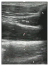
Ultrasound view of the subclavius muscle. SM: subclavius muscle, C: clavicle, V: subclavian vein and/or infraclavicular axillary vein, R1: first rib, R2: second rib, and L: lung.

References

    1. Aitken D. R., Minton J. P. The ‘pinch-off sign’: a warning of impending problems with permanent subclavian catheters. The American Journal of Surgery. 1984;148(5):633–636. doi: 10.1016/0002-9610(84)90340-4. - DOI - PubMed
    1. Hinke D. H., Zandt-Stastny D. A., Gooodman L. R., Quebbeman E. J., Krzywda E. A., Andris D. A. Pinch-off syndrome: a complication of implantable subclavian venous access devices. Radiology. 1990;177(2):353–356. doi: 10.1148/radiology.177.2.2217768. - DOI - PubMed
    1. Magney J. E., Flynn D. M., Parsons J. A., et al. Anatomical mechanisms explaining damage to pacemaker leads, defibrillator leads, and failure of central venous catheters adjacent to the sternoclavicular joint. Pacing and Clinical Electrophysiology. 1993;16(3, part 1):445–457. doi: 10.1111/j.1540-8159.1993.tb01607.x. - DOI - PubMed
    1. Krutchen A. E., Bjarnason H., Stackhouse D. J., Nazarian G. K., Magney J. E., Hunter D. W. The mechanisms of positional dysfunction of subclavian venous catheters. Radiology. 1996;200(1):159–163. doi: 10.1148/radiology.200.1.8657904. - DOI - PubMed
    1. Franey T., DeMarco L. C., Geiss A. C., Ward R. J. Catheter fracture and embolization in a totally implanted venous access catheter. Journal of Parenteral and Enteral Nutrition. 1988;12(5):528–530. doi: 10.1177/0148607188012005528. - DOI - PubMed

LinkOut - more resources