. 2016:2016:6810980.
doi: 10.1155/2016/6810980. Epub 2016 Feb 4.

Standardizing Umbilical Cord Mesenchymal Stromal Cells for Translation to Clinical Use: Selection of GMP-Compliant Medium and a Simplified Isolation Method

Affiliations

Standardizing Umbilical Cord Mesenchymal Stromal Cells for Translation to Clinical Use: Selection of GMP-Compliant Medium and a Simplified Isolation Method

J Robert Smith et al. Stem Cells Int. 2016.

Abstract

Umbilical cord derived mesenchymal stromal cells (UC-MSCs) are a focus for clinical translation but standardized methods for isolation and expansion are lacking. Previously we published isolation and expansion methods for UC-MSCs which presented challenges when considering good manufacturing practices (GMP) for clinical translation. Here, a new and more standardized method for isolation and expansion of UC-MSCs is described. The new method eliminates dissection of blood vessels and uses a closed-vessel dissociation following enzymatic digestion which reduces contamination risk and manipulation time. The new method produced >10 times more cells per cm of UC than our previous method. When biographical variables were compared, more UC-MSCs per gram were isolated after vaginal birth compared to Caesarian-section births, an unexpected result. UC-MSCs were expanded in medium enriched with 2%, 5%, or 10% pooled human platelet lysate (HPL) eliminating the xenogeneic serum components. When the HPL concentrations were compared, media supplemented with 10% HPL had the highest growth rate, smallest cells, and the most viable cells at passage. UC-MSCs grown in 10% HPL had surface marker expression typical of MSCs, high colony forming efficiency, and could undergo trilineage differentiation. The new protocol standardizes manufacturing of UC-MSCs and enables clinical translation.


Figures

Figure 1
Figure 1
A schematic of the optimized isolation method. The major steps: (a) umbilical cord selected. (b) 1 cm section prior to cutting into 4 equal pieces. (c) Cord pieces rinsed in DPBS. (d) The cord pieces inside a C-tube immersed in enzyme solution. (e) Dissociation with C-tubes and Miltenyi Dissociator. (f) Steps following dissociation prior to plating the isolated cells. (g) The isolated cell initial plating at P0 and subsequent expansion over multiple passages.
Figure 2
Figure 2
Effect of various experimental variables on UC-MSCs isolation. (a) The new methods average cell number per cm of umbilical cord isolated, compared to the old cell number isolated per cm (∗∗ means p < 0.001). (b) Comparing different experimental variables. Significant difference observed in Caesarean-section delivery versus vaginal delivery ( means p < 0.05). (c) Population doubling time for passage 0 (initial isolation) or passage 1 (first passage of expansion phase). represents p < 0.05 for 2% hpl media compared to 5% and 10% media. † represents p < 0.05 for the passages (P0 compared to the P1).
Figure 3
Figure 3
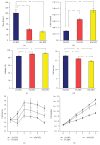
Effect of HPL concentration on expansion. (a–d) UC-MSC (n = 6) expansion results combined for passages 1–5. (a) Population doubling times for the 3 media conditions. (b) Number of cells counted at passage for each media condition. (c) Cell viability at passage for each media condition. (d) The average size of the cell for each media condition at passage. (e) Cell size over 5 passages for each media condition. (f) The theoretical yield if an entire umbilical cord was isolated and grown to confluence at each passage. means p < 0.05 and ∗∗ means p < 0.001.
Figure 4
Figure 4
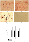
Differentiation and colony forming unit fibroblast (CFU-F) results for the characterization of UC-MSCs. (a) After adipogenic differentiation, MSCs were stained with Oil Red which binds to lipid droplets (20x objective magnification; scale bar = 200 micrometers). (b) After osteogenic differentiation, MSCs were stained with Alizarin Red S which binds to calcium deposits. (c) After chondrogenic differentiation, MSCs stained with Safranin O which binds to glycosaminoglycans in cartilage ((b) and (c) at 10x objective magnification; scale bar = 400 micrometers). (d) UC-MSCs in normal growth conditions (control) phase contrast micrograph at 4x objective magnification. (e) CFU-F efficiency was calculated by dividing the number of plated cells by the number of CFU-F colonies observed. Panel (e) shows colony forming efficiency versus human pooled platelet lysate (HPL) concentration in medium (2, 5, or 10% HPL) after plating at 5 (black bars) or 10 (gray bars) cells per cm2 and 4 days in culture.
Figure 5
Figure 5
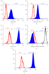
Histograms of flow cytometry; blue solid filled overlay represents the test sample; red diagonal line filled overlay represents the isotype control. For each histogram the negative gate (red bar) was set for inclusion of 99% of the isotype. Percentages shown in histograms are for the test samples. (a–c) All are markers in the positive cocktail, CD90, CD105, and CD73. The positive gate percentages shown in blue for each sample. (d) The negative cocktail, with CD44 included as a positive control (unfilled overlay) positive percentage in black. (e) CD44 marker included outside of the positive cocktail.

References

    1. Sensebé L., Gadelorge M., Fleury-Cappellesso S. Production of mesenchymal stromal/stem cells according to good manufacturing practices: a review. Stem Cell Research and Therapy. 2013;4(3, article 66) doi: 10.1186/scrt217. - DOI - PMC - PubMed
    1. Mendicino M., Bailey A. M., Wonnacott K., Puri R. K., Bauer S. R. MSC-based product characterization for clinical trials: an FDA perspective. Cell Stem Cell. 2014;14(2):141–145. doi: 10.1016/j.stem.2014.01.013. - DOI - PubMed
    1. Krampera M., Galipeau J., Shi Y., Tarte K., Sensebe L. Immunological characterization of multipotent mesenchymal stromal cells—the International Society for Cellular Therapy (ISCT) working proposal. Cytotherapy. 2013;15(9):1054–1061. doi: 10.1016/j.jcyt.2013.02.010. - DOI - PubMed
    1. Dominici M., Le Blanc K., Mueller I., et al. Minimal criteria for defining multipotent mesenchymal stromal cells. The International Society for Cellular Therapy position statement. Cytotherapy. 2006;8(4):315–317. doi: 10.1080/14653240600855905. - DOI - PubMed
    1. Ringdén O., Erkers T., Nava S., et al. Fetal membrane cells for treatment of steroid-refractory acute graft-versus-host disease. STEM CELLS. 2013;31(3):592–601. doi: 10.1002/stem.1314. - DOI - PubMed